


Hybrid Imaging Modalities: Combining Functional and Anatomical Insights to Optimize Diagnostics and Treatment
The evolution of medical imaging has reached a transformative milestone with the development of hybrid imaging modalities that seamlessly integrate functional and anatomical information within a single examination. These sophisticated systems represent a paradigm shift from traditional single-modality approaches, delivering comprehensive diagnostic insights that neither functional nor anatomical imaging could achieve independently. The global hybrid imaging market, valued at approximately $8.2 billion in 2023, continues to expand as healthcare providers recognize the profound impact these technologies have on diagnostic accuracy, treatment planning, and patient outcomes.
Modern hybrid imaging modalities have fundamentally altered the landscape of medical diagnostics by addressing the inherent limitations of standalone imaging techniques. Where conventional anatomical imaging provides structural detail but lacks information about physiological processes, and functional imaging reveals metabolic activity without precise anatomical localization, hybrid systems create synergistic combinations that enhance diagnostic capabilities far beyond the sum of their individual components. This integration has proven particularly valuable in complex clinical scenarios where precise anatomical correlation of functional abnormalities is essential for accurate diagnosis and optimal treatment planning.
The Foundation of Hybrid Imaging Excellence
Technical Integration and System Architecture
The development of hybrid imaging modalities required overcoming substantial technical challenges related to hardware integration, data acquisition synchronization, and image co-registration. Positron Emission Tomography combined with Computed Tomography represents the most established hybrid technology, with over 7,000 PET/CT systems operating worldwide as of 2024. The sequential acquisition approach employed in PET/CT systems allows for rapid CT scanning followed by PET imaging, enabling precise anatomical localization of metabolic abnormalities while providing attenuation correction for quantitative PET analysis.
Single Photon Emission Computed Tomography integrated with CT has emerged as another highly successful hybrid modality, particularly valuable in clinical scenarios where radiopharmaceutical localization requires anatomical context. SPECT/CT systems have demonstrated remarkable versatility across multiple medical specialties, with studies consistently showing diagnostic accuracy improvements of 15-20% compared to SPECT alone. The technology has proven especially beneficial in bone imaging, where the combination of functional bone metabolism information with detailed skeletal anatomy enhances detection and characterization of both benign and malignant conditions.
The newest addition to the hybrid imaging family, PET/MRI, represents the most technologically sophisticated approach, combining the metabolic insights of PET with the superior soft tissue contrast and functional capabilities of magnetic resonance imaging. These systems offer unique advantages in pediatric imaging and neurological applications, where the reduced radiation exposure of MRI compared to CT becomes particularly significant. However, the complexity of simultaneous PET and MRI acquisition has required innovative solutions for attenuation correction and magnetic field interactions that continue to evolve.
Radiopharmaceutical Integration and Multimodal Contrast Agents
The success of hybrid imaging modalities depends critically on the availability of appropriate radiopharmaceuticals and contrast agents that can provide meaningful functional information while maintaining compatibility with anatomical imaging requirements. Fluorine-18 fluorodeoxyglucose remains the most widely utilized PET tracer, providing information about glucose metabolism that proves invaluable in oncology, cardiology, and neurology applications. The 110-minute half-life of F-18 allows sufficient time for comprehensive hybrid imaging protocols while minimizing radiation exposure concerns.
Emerging radiopharmaceuticals designed specifically for hybrid imaging applications are expanding the diagnostic capabilities of these systems. Gallium-68 labeled compounds have gained prominence for neuroendocrine tumor imaging, while newer tracers targeting specific receptors and cellular processes continue to broaden the clinical applications of hybrid imaging. The development of multimodal contrast agents that can be detected by both PET and MRI components represents an active area of research that promises to further enhance the synergistic benefits of hybrid systems.
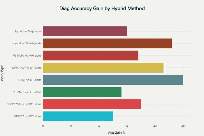
Diagnostic accuracy improvements achieved through hybrid imaging modalities compared to single-modality approaches
Clinical Applications and Diagnostic Impact
Oncological Excellence Through Integrated Imaging
In oncology, hybrid imaging modalities have fundamentally transformed cancer diagnosis, staging, and treatment monitoring. PET/CT has achieved widespread adoption as the gold standard for cancer staging, with diagnostic accuracy rates consistently exceeding 92% in lung cancer, lymphoma, and melanoma applications. The ability to simultaneously assess tumor metabolism and anatomical extent provides oncologists with comprehensive information essential for treatment planning, from initial staging through response monitoring and surveillance for recurrence.
The integration of functional and anatomical information has proven particularly valuable in distinguishing between treatment-related inflammation and residual or recurrent tumor tissue. This capability has significant implications for patient management, as it enables clinicians to avoid unnecessary interventions when apparent abnormalities represent benign post-treatment changes rather than active disease. Studies demonstrate that hybrid imaging reduces equivocal interpretations by up to 40% compared to single-modality approaches, leading to more confident clinical decision-making and improved patient outcomes.
SPECT/CT applications in oncology focus primarily on specialized scenarios such as sentinel lymph node mapping, neuroendocrine tumor localization, and radioiodine therapy planning for thyroid cancer. The technology has shown particular strength in bone metastasis detection, where the combination of radiopharmaceutical bone uptake with detailed skeletal anatomy provides superior sensitivity and specificity compared to conventional bone scintigraphy. This enhanced performance directly impacts treatment decisions, enabling more accurate staging and appropriate selection of therapeutic interventions.
Cardiovascular Applications and Risk Stratification
Cardiac applications of hybrid imaging modalities have revolutionized the assessment of coronary artery disease and myocardial viability. SPECT/CT myocardial perfusion imaging combines functional assessment of coronary blood flow with anatomical information about cardiac structure and coronary calcification, providing comprehensive evaluation that guides both medical and interventional treatment decisions. The technology enables clinicians to correlate perfusion defects with specific coronary territories and assess the likelihood of successful revascularization procedures.
The integration of CT-based attenuation correction in cardiac SPECT/CT has significantly reduced artifacts that historically complicated interpretation of myocardial perfusion studies, particularly in obese patients and those with breast attenuation. This improvement has enhanced diagnostic confidence and reduced the need for repeat studies or additional imaging modalities. Clinical studies demonstrate that hybrid cardiac imaging influences treatment decisions in approximately 30-40% of patients compared to single-modality approaches.
PET/MRI cardiac applications focus primarily on assessment of myocardial viability and evaluation of cardiac sarcoidosis and other inflammatory conditions. The superior soft tissue contrast of MRI combined with the metabolic information from PET provides detailed assessment of myocardial tissue characteristics that cannot be achieved with other imaging modalities. This information proves particularly valuable in patients being considered for cardiac transplantation or high-risk revascularization procedures.
Neurological and Brain Imaging Advancements
In neurology and psychiatry, hybrid imaging modalities have enhanced the diagnosis and monitoring of neurodegenerative diseases, brain tumors, and epilepsy. PET/CT brain imaging provides valuable information about glucose metabolism and specific receptor binding that correlates with cognitive function and disease progression. The technology has proven particularly valuable in differentiating between various forms of dementia and in monitoring treatment response in brain tumor patients.
PET/MRI has emerged as the preferred hybrid modality for many neurological applications due to the superior soft tissue contrast of MRI and the reduced radiation exposure compared to CT. The technology enables simultaneous assessment of brain structure, function, and metabolism, providing comprehensive information that supports both diagnosis and treatment planning. Studies indicate that PET/MRI detects clinically significant findings missed by PET/CT in more than 50% of brain tumor patients, highlighting the importance of optimal soft tissue contrast in neurological imaging.
The application of hybrid imaging in epilepsy evaluation has transformed surgical planning by providing precise localization of epileptogenic foci relative to critical brain structures. This information enables neurosurgeons to plan more targeted resections while minimizing risks to eloquent brain regions. The technology has contributed to improved surgical outcomes and reduced morbidity in patients with medically refractory epilepsy.
Technological Advantages and Clinical Benefits
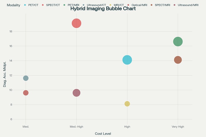 Cost-effectiveness analysis of hybrid imaging modalities showing relationship between cost, accuracy improvement, and clinical applications
Quantitative Analysis and Standardization
Hybrid imaging modalities provide unique opportunities for quantitative analysis that enhance diagnostic accuracy and enable standardized interpretation across different institutions and imaging systems. The integration of CT-based attenuation correction in both PET/CT and SPECT/CT enables accurate quantification of radiopharmaceutical uptake, supporting more precise diagnosis and improved monitoring of treatment response. This quantitative capability has proven particularly valuable in oncology applications where standardized uptake values provide objective measures of treatment response.
The anatomical information provided by hybrid systems enables more accurate region-of-interest analysis by providing precise boundaries for functional measurements. This capability reduces inter-observer variability and improves the reproducibility of quantitative assessments. Clinical studies demonstrate that standardized quantitative analysis of hybrid imaging data correlates more strongly with clinical outcomes compared to qualitative interpretation alone.
Advances in artificial intelligence and machine learning are further enhancing the quantitative capabilities of hybrid imaging systems. Automated analysis algorithms can extract complex features from both functional and anatomical components of hybrid images, providing insights that may not be apparent to human interpreters. These developments promise to further improve diagnostic accuracy while reducing interpretation time and inter-observer variability.
Workflow Efficiency and Patient Experience
The integration of multiple imaging modalities into single examination sessions provides significant benefits for both healthcare providers and patients. Hybrid imaging reduces the need for separate appointments and multiple imaging studies, improving workflow efficiency and reducing healthcare costs. Patients benefit from reduced travel time, fewer appointments, and more convenient scheduling, leading to improved satisfaction and adherence to imaging recommendations.
The reduction in total examination time achieved through hybrid imaging also decreases patient anxiety and discomfort associated with prolonged medical procedures. This benefit proves particularly important in pediatric applications and in patients with claustrophobia or other conditions that make prolonged imaging challenging. Studies indicate that patient satisfaction scores are consistently higher for hybrid imaging procedures compared to sequential single-modality examinations.
Healthcare providers benefit from more efficient utilization of imaging resources and reduced scheduling complexity. The comprehensive information provided by hybrid imaging often eliminates the need for additional imaging studies, reducing both costs and radiation exposure. This efficiency enables healthcare systems to serve more patients while maintaining high diagnostic standards.
Future Directions and Emerging Technologies
The continued evolution of hybrid imaging modalities promises even greater integration of functional and anatomical information with enhanced diagnostic capabilities. Artificial intelligence integration is expected to revolutionize image analysis and interpretation, providing automated detection of abnormalities and quantitative analysis that enhances diagnostic accuracy while reducing interpretation time. These developments will democratize access to expert-level image interpretation and reduce healthcare disparities related to imaging expertise availability.
Novel radiopharmaceuticals designed specifically for hybrid imaging applications continue to expand the clinical capabilities of these systems. Theranostic approaches that combine diagnostic imaging with targeted therapy are emerging as particularly promising applications of hybrid imaging technology. These developments enable personalized treatment approaches based on individual tumor characteristics and treatment response patterns.
The integration of hybrid imaging with other advanced technologies such as artificial intelligence, robotics, and precision medicine platforms promises to create comprehensive diagnostic and treatment systems that optimize patient outcomes while minimizing healthcare costs. These developments represent the future of personalized medicine, where individual patient characteristics guide both diagnostic and therapeutic decisions.
Modern hybrid imaging modalities have established themselves as indispensable tools in contemporary medical practice, providing diagnostic capabilities that significantly exceed those of single-modality approaches. The integration of functional and anatomical information within single examination sessions has transformed clinical practice across multiple specialties while improving patient outcomes and healthcare efficiency. As technology continues to advance and new applications emerge, hybrid imaging modalities will play an increasingly central role in delivering precision medicine that optimizes diagnostic accuracy while minimizing patient burden and healthcare costs.

