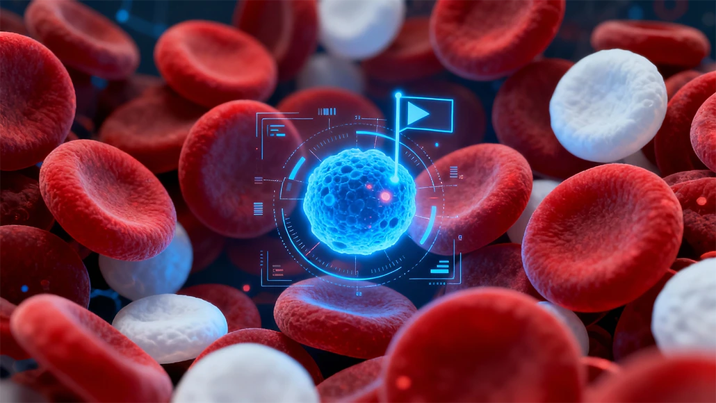
An AI tool that can evaluate the abnormal blood cells, and with greater precision and reliability as compared to human experts, could as well change the way conditions like leukemia get diagnosed.
Researchers have gone ahead and created a system named CytoDiffusion, which makes use of generative AI, which is the same kind of technology behind the image generators like DALL-E, in order to study the shape as well as the structure of blood cells.
Unlike the numerous other AI models, which are trained to simply recognize the patterns, CytoDiffusion, which is developed by the researchers at the University of Cambridge, University College London as well as Queen Mary University of London, could precisely pinpoint a wide range of normal blood cell appearances and also spot unusual or rare cells, which may indicate the disease. The results are indicated in the Nature Machine Intelligence journal.
Apparently, the spotting of subtle differences within the blood cell size and shape as well as the appearance is indeed a cornerstone when it comes to diagnosing numerous blood disorders. However, the task needs to have years of training, and even then, there would be varied doctors who can disagree on challenging cases. According to the study’s first author from the Department of Applied Mathematics and Theoretical Physics in Cambridge, Simon Deltadahl, all got numerous different kinds of blood cells, which have varied properties as well as different roles within the body. There are white blood cells that specialize in fighting the infection, for instance. Knowing what an unusual or diseased blood cell would look like under a microscope is indeed an important part of diagnosing numerous diseases.
But a typical blood smear goes on to have thousands of cells, which are far more than any human could evaluate. Deltadahl adds that humans cannot have a look at all the cells in a smear, as it is just not possible. Their model can go ahead and automate that process, highlight anything that looks unusual for human review, and triage the routine cases.
As per co-senior author from Queen Mary University of London, Dr. Suthesh Sivapalaratnam, the clinical challenge he went on to face as a junior hematology doctor was that after a day of work, he would have a lot of blood films to evaluate. As he was evaluating them in the late hours, he became convinced that AI would surely do a much better job than himself.
In order to develop CytoDiffusion, the researchers went on to train the system on more than half a million images of blood smears that were collected at Addenbrooke’s Hospital located in Cambridge. The dataset, which is the largest of its kind, had both common blood cell types as well as rarer examples and also elements that can confuse the automated systems. Through modelling the complete distribution of cell appearances and not just learning to separate the categories, the AI became much more robust to differences between the hospitals, microscopes, and staining methods, and also better able to gauge the rare or abnormal blood cells.
In tests, CytoDiffusion could as well detect abnormal blood cells, which are linked to leukemia, with far more sensitivity as compared to the existing systems. It also matched or even at times surpassed the present state-of-the-art models, even when given far fewer training examples, and also quantified its own uncertainty. Deltadahl added that when they tested its precision, the system was slightly better as compared to humans; however, where it actually stood out was in knowing when it was not sure. Their model would never say that it was certain and then be wrong; however, that is something that the humans sometimes do.
Professor Michael Roberts, the co-senior author, also from the Department of Applied Mathematics and Theoretical Physics of Cambridge, said that they assessed the method against many of the issues that were seen in machines and also the degree of uncertainty within the labels. This framework goes on to give a multi-faceted view of the model performance that they believe is indeed going to be beneficial to the researchers.
In addition to this, the team also showed that CytoDiffusion could also generate synthetic blood cell images that are indistinguishable from real ones. In one of the Turing tests with ten experienced hematologists, human experts were indeed no better than having a chance at telling the real from the AI-generated images. Deltadahl recalled that really surprised him. These are people who go on to stare at blood cells all times of the day, and even they could not tell.
As part of the project, the researchers are also releasing what, according to them, is the largest publicly available dataset pertaining to peripheral blood smear images in the world, which is more than half a million overall. Through making this resource open, they also hope to empower the researchers across the world so as to build as well as test new AI models, democratize the access when it comes to high-quality medical data, and, at the end of the day, contribute to much better patient care.
While the results really look quite promising, the researchers go on to say that CytoDiffusion isn’t a replacement for the trained clinicians. Rather, it is designed in order to support them by accelerating the flagging of abnormal cases for review and also handling many more routine ones in an automated form. UCL’s Professor Parashkev Nachev, who is the co-senior author, says that the true value of healthcare AI lies not just in approximating the human expertise at a lower cost, but rather in enabling greater diagnostic, prescriptive, and prognostic power than what either the experts or simple statistical models can attain. Their work suggests that generative AI is indeed going to be central to this mission, transforming not just the fidelity when it comes to clinical support systems but also their insight into limits pertaining to their own knowledge.
This kind of metacognitive awareness, knowing what one does not know, is crucial to clinical decision-making, and there are machines that may be better at it as opposed to what humans are.
According to the researchers, further work is required so as to make the system much faster and to also test it throughout the diverse patient populations in order to make sure of fairness as well as precision.
Notably, the research was supported in part by Wellcome, the British Heart Foundation, the Trinity Challenge, the NHS Foundation Trust of the Cambridge University Hospitals, Barts Health NHS Trust, the NIHR UCLH Biomedical Research Centre, the NIHR Cambridge Biomedical Research Centre, and the NHS Blood and Transplant. The research was conducted through the imaging working group of the BloodCounts! consortium, which looks forward to using AI in order to improve the blood diagnostics across the world.


