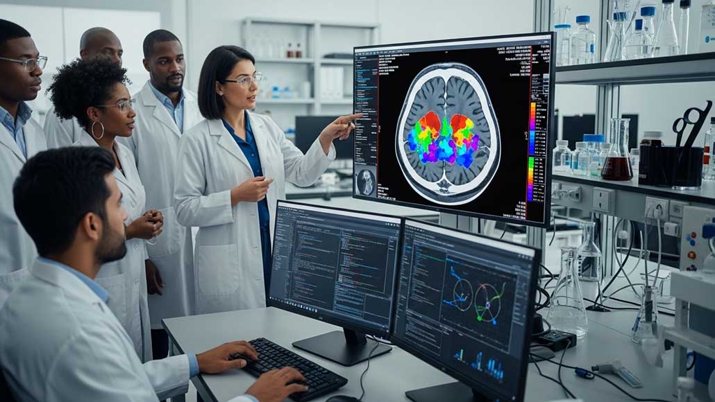
A new artificial intelligence tool has cropped up that can make it much easier and cheaper for doctors as well as researchers to train medical imaging software, even at a time when only a small number of patient scans are available. This AI tool enhances a process named medical image segmentation wherein every pixel in the image is labelled based on what it happens to represent—normal or cancerous tissue, for instance. This kind of process is often performed by way of highly trained experts, and its deep learning has shown promise when it comes to automating this kind of labor-intensive task.
However, the bigger challenge is that deep learning-based methods happen to be data hungry, which require massive amounts of pixel-by-pixel annotated images in order to learn, says Lee Zhang, a student in the department of electrical and computer engineering at the University of California in San Diego, who also has a PhD.
So as to overcome this limitation, Zhang, along with his team of researchers, which is led by Professor Pengtao Xie, the UC San Diego electrical and computer engineering professor, has gone on to develop an AI tool that can learn the image segmentation from a very small number of expert-labeled samples. By way of doing so, it happens to cut the amount of data that is usually needed by almost 20 times. All this could potentially lead to much faster, more affordable diagnostic tools, specifically within clinics as well as hospitals having limited resources.
Apparently, the AI tool was tested on a range of medical image segmentation tasks. It went on to gauge and identify skin lesions in dermoscopy images, breast cancer in ultrasound scans, and placental vessels in fetoscopic images, along with polyps in colonoscopy images and also foot ulcers within standard camera photos, just to cite a few examples. This method was also extended to certain 3-D images – those that are used to map the hippocampus or liver.
In scenarios where annotated data went on to be extremely limited, the AI tool boosted the model performance between 10 and 20% as against the present approach. It needed 8 to 20 times less real-world training data as compared to standard methods while at the same time matching or even outperforming them.
Zhang went on to describe how this AI tool could potentially be made use of in order to help dermatologists diagnose skin cancer. Rather than gathering and labeling thousands of images, a trained expert within the clinic might only require annotating 40, for instance. The AI tool could use this very small dataset in order to identify any suspicious lesions from the dermoscopy images of a patient, and that too in real time. As per Zhang, it could very well help the doctors to make a faster and more precise diagnosis.
It is well to be noted that the system works in stages. First, it happens to learn how to generate synthetic images coming from segmentation masks, which are indeed necessary for color-coded overlays that happen to tell an algorithm which parts of the image are healthy or diseased. Then it makes use of that knowledge to create a novel and artificial image – mask pairs—in order to speed up a small data set of real examples. A segmentation model is trained by way of using both. Through a consistent feedback loop, the system then refines the images that it creates based on how well they enhance the learning of the model.
According to Zhang, the feedback loop happens to be a big part of what makes the system work very well. He adds that rather than treating the data generation along with segmentation model training as two different tasks, this system happens to be the first to put them together. The segmentation performance goes on to guide the data generation process. This kind of makes sure that the synthetic data is not just realistic, but at the same time, it is specifically customized in order to make sure that the model segmentation capabilities are improved.
Marching forward, the team looks forward to making their AI tools more versatile as well as smarter. The researchers are also looking forward to incorporating feedback from clinicians directly within the training process so as to make the generated data even more relevant when it comes to real-world medical usage.


