During the past three decades, investment, innovation and advances in new technologies and procedures have markedly improved our ability to detect and diagnose early stage breast cancer and have had an enormous positive impact on patients. However, improvements in breast localization procedures and technologies have lagged, and many hospitals continue to rely on wire localization procedures to guide surgeons to breast lesions on the day of surgery.
With new advances in wire-free localization technologies for surgical guidance and excision of small, non-palpable, imaging-detected malignant or high-risk breast lesions, Mills Peninsula Medical Center saw an opportunity to improve upon the quality of care and patient experience for those requiring these procedures, while improving efficiency and capacity for these services and procedures.
Harriett Borofsky, MD, Medical Director of Women Imaging, explains how Mills-Peninsula Medical Center implemented wire-free localization.
What was the main objective in implementing wire-free localization?
In embarking upon wireless localization procedures, our objectives were to assess overall patient tolerance and satisfaction while undergoing these procedures, to determine ease and accuracy of reflector placements and complications for the full gamut of mammographic, tomographic and ultrasound detected lesions, including those lesions requiring bracketing, and to assure successful intra-operative removal of all reflectors and index lesions. Secondary outcomes would be improved overall efficiency and capacity of radiology and operating room services and positive impact on margin status, compared to traditional wire localization procedures.
 Who were the champions of this effort?
The wire-free localization effort was led by myself and Andrea Metkus, M.D., Medical Director of the Breast Cancer Program and breast surgeon at Mills-Peninsula Medical Center.
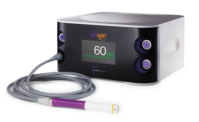
After a review of available wire-free localization technologies, the Radar Localization System was selected for a 15-patient feasibility study. The lead radiologist was trained in reflector placement and deployment and the lead surgeon was trained in intra-operative reflector retrieval procedures. All patients diagnosed, via minimally invasive tomographic, stereotactic, ultrasound or MRI-guided core needle biopsies, with high-risk lesions requiring surgical excision or malignant lesions amenable to breast conservation surgery with lumpectomy, were referred for surgical consultation.The surgeon then referred appropriate patients for reflector placement prior to their scheduled surgery. The first 15 cases were performed and reviewed for successful reflector placement and retrieval, complications and patient tolerance and satisfaction.
Our feasibility study and results were officially presented to the New Procedure and Technology Evaluation Committee, Mills-Peninsula Medical Center, comprised of the hospital’s CEO, CFO, Chief-of-Staff, Vice Chief-of-Staff, Nursing Supervisor, managers of the radiology and surgical departments and foundation president.
Following official approval, the wireless technology was funded by the Mills-Peninsula Medical Center Foundation. The new technology was then promoted and communicated to the hospital staff and physicians through department meetings and a written article in the Professional Medical Staff Newsletter.
What outcomes were observed upon completion of the project?
Our project and feasibility study on wire-free localization procedures confirmed a positive impact on patient experience for those requiring these procedures, an improved overall efficiency and capacity for the radiology and OR departments by decoupling the surgery and radiology schedules, successful localization and surgical excision of reflectors and index lesions in all cases and elimination of surgical mishaps related to wires and wire localization procedures.
For many patients, the most dreaded aspect of breast surgery is the needle localization prior to the procedure. This requires patients, who are NPO, to have uncomfortable wires placed two hours prior to scheduled surgery. Wire-free localization procedures proved to be easily and conveniently scheduled, well tolerated by all patients and without complications.
All index lesions, including those requiring bracketing, were able to be accurately localized. Our experience confirmed this procedure to be easily learned, quickly performed and incorporated into the breast imaging center’s workflow for all localizations, including bracketing of complex lesions, with high accuracy.
For the operating room schedule, wire-free localization increased capacity to start procedures early in the morning, while eliminating unexpected delays due to vasovagal events, complications and transportation issues related to wire localizations on the day of surgery. For the radiology/breast imaging department, the technology eliminated the need to place wires on the day of surgery and avoided procedure and capacity conflicts.
How did your team innovate to reach the stated goals of the project?
This was the first collaborative project conducted between the breast center and the outpatient OR teams, supported by the administration and funded by the foundation. We had several meetings of lead physicians, staff and administration to discuss the project, address concerns and training and assure seamless workflow and patient safety. It was an exciting collaborative effort, benefitting our mutual patients and our department’s workflow, which was embraced by all.
To achieve a successful wire-free program, it was essential to create new protocols and work flows for wire-free localization scheduling, procedure performance, image access on the day procedure and specimen radiography and documentation on the day of procedure. This too was a collaborative effort between surgery and radiology scheduling and OR staff, requiring changing decades-old habits.
While we are still refining our processes for wire-free localizations, we are so encouraged by the positive impact it has had on overall patient experience and quality of care, easing patient anxiety and procedure time on the day of surgery, while improving workflow and overall capacity, especially in the operating room.


