


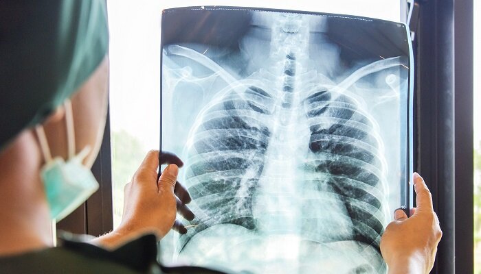
According to new research, if modified for use in hospitals and clinics, an imaging technology that is predominantly used in research labs could detect early-stage lung disease.
Researchers at the KTH Royal Institute of Technology in Stockholm investigated how phase-contrast X-ray imaging could be used to detect lung illness in humans. A model of the human chest created by Duke University was used by the research team in the study that was titled- Phase-contrast virtual chest radiography.
The researchers discovered that phase-contrast chest radiography could see the smallest airways in the lungs, including some as small as 2 mm broad. The method can also aid in the detection of lung disease-related blockages, which conventional radiography cannot.
The conventional imaging limitations
In research labs, conventional phase-contrast imaging is limited to imaging centimeter-scale soft tissue samples. This study demonstrated that, if the technological requirements for clinical application can be met, phase-contrast X-ray imaging has the potential to identify lung disease.
According to Ilian Haggmark, the study’s primary author, conventional chest radiography technologies used in clinics and hospitals today can help detect lung disease, but they are limited by how they generate images. Haggmark opines that phase-contrast X-ray imaging may extract more information at a higher resolution while using the same amount of radiation exposure as traditional radiography.
In traditional radiography, the X-ray beam goes through the body and is absorbed in varying amounts by different tissues. After passing through the body, a detector assesses the intensity of the beam. This is known as attenuation, and it is the fundamental mechanism that provides the contrast that generates X-ray images.
Clinicians were able to obtain more information from each X-ray beam using the phase-contrast technique. They can use this technology to analyse changes in the waveforms of X-rays passing through a sample. X-ray beams collide with atoms and other structures, which can cause a wave’s location to change at any point concerning a reference wave.
The use of phase-contrast technology aids in the detection of lung disease
This data is utilised to create an image that enriches the sample’s structures. With enhanced contrast and resolution, these pictures can emphasise the margins of bronchial walls and tiny airways, allowing clinicians to quickly recognise the symptoms of lung disease.
Moving the detector farther from the patient, according to the researchers, is critical to this strategy. They also stress that building imaging equipment for larger samples will take time. A high-power X-ray source with a tiny emission spot is required. Essentially, bright X-ray sources are required, according to Haggmark.
As per the experts, interesting developments are occurring, but it will take time before they are ready for human usage. For the time being, simulations as well as virtual clinical trials are ideal tools for exploring what one can achieve once the source technology is ready, according to Haggmark.

