Close
Digital Health & Ai Innovation summit 2026
Medical Taiwan 2026

UPMC to Expand Telehealth Network in Italy to Battle COVID-19 Surge

UPMC to Expand Telehealth Network in Italy to Battle COVID-19 Surge

UPMC has been awarded almost $2 million to launch a telemedicine platform in Italy, which was hit hard by the coronavirus pandemic earlier this year and is now facing another wave.

The United States Agency for International Development (USAID) is giving a $1.8 million grant to UPMC in Italy, part of the Pittsburgh-based health systemโ€™s international network, to create a telehealth program aimed at serving the nationโ€™s frailest patients and reducing congestion in its health systems and hospitals.

โ€œThanks to the support of the US Government, UPMC in Italy will be able to build an integrated telemedicine platform based on the state-of-the-art practices we have developed over the course of 20 years in Italy,โ€ Bruno Gridelli, executive vice president of UPMC International and country manager of UPMC in Italy, said in a press release. โ€œThe Italian health care system will benefit from an innovative tool that helps to manage this emergency and improves the flexibility of and access to health care services long term.โ€

UPMCโ€™s international arm supports healthcare networks in Italy, Ireland, China and Kazakhstan. In Italy, that network is focused on seven sites.

One of the hardest-hit countries in Europe, Italy has seen more than 790,000 cases of COVID-19 and lost close to 40,000 residents, with daily cases and deaths jumping 50 percent over the past week. The country has the second highest death toll behind the UK.

Last week, the nation listed several regions as โ€œred zones,โ€ and governmental leaders urged the public to practice caution to avoid new lockdowns that are being implemented in England, France and Germany. People in those red zones โ€“ which include Milan and the northern regions of Lombardy and Piedmont โ€“ will only be allowed to leave their homes for work, health needs or emergencies, while all schools have moved to online learning.

UPMC, which has an extensive connected health network in the US, is expected to use that expertise to help Italyโ€™s beleaguered health system. Telehealth programs will be up and running on a trial basis through the first half of 2021.

โ€œThe pandemic has brought telemedicine to the forefront of health care, presenting us with an opportunity to protect the most high-risk patients, reduce costs, collect useful data and treat patients in the comfort of their homes,โ€ Gridelli said in the press release. โ€œLooking beyond the pandemic, this platform will create increasingly immediate and personalized health care in Italy.โ€


Cerapedics Announces Commercial Launch of i-FACTOR Matrix in Canada

   Cerapedics Announces Commercial Launch of i-FACTOR Matrix in Canada

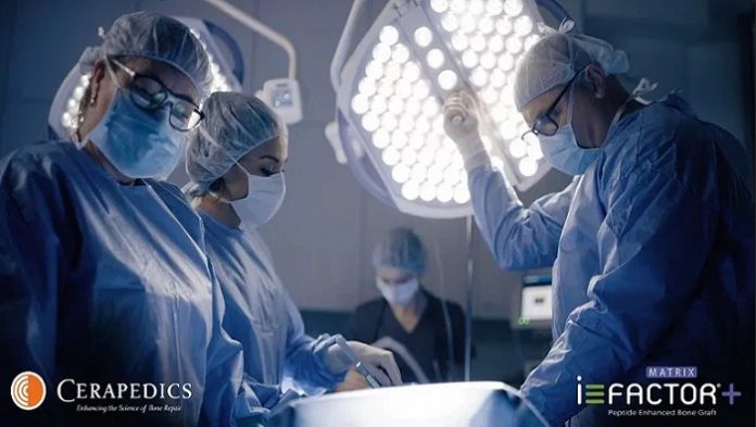
Cerapedics, a private ortho-biologics company, today announced the full commercial availability of i-FACTOR+ MATRIX for surgical implantation in Canada.

โ€œWe are excited to announce that i-FACTOR+ Matrix is now fully available in Canada through our distributor Surgi-One and can be used in both spine and general orthopedic applications,โ€ said Glen Kashuba, Chief Executive Officer of Cerapedics, โ€œCanada will be the first market offering the i-FACTOR+ MATRIX product, which leverages the clinically proven P-15 osteogenic cell-binding peptide combined with a collagen carrier for optimized handling.โ€

i-FACTOR+ MATRIX utilizes a small proprietary peptide (P-15) technology developed by Cerapedics to support bone growth through cell attraction, attachment, and activation. Cerapedics initial P-15 technology product, i-FACTOR Peptide Enhanced Bone Graft, was approved in the European Union in 2008 and Australia in 2010 for use in the repair of bony voids or defects in orthopedic applications throughout the skeletal system (i.e., the spine and extremities). In the United States, i-FACTOR Peptide Enhanced Bone Graft was approved via a U.S. Food and Drug Administration (FDA) premarket approval (PMA) application in 2015 only for use in single-level anterior cervical fusion procedures.

โ€œIn addition to the commercial launch in Canada, the latest i-FACTOR+ MATRIX generation is currently being studied in our FDA IDE study, ASPIRE, targeting lumbar fusions (TLIF), as previously announced. i-FACTOR+ Matrix is the same technology as the P15-L investigational material in the IDE,โ€ said Jeffrey Marx, Ph.D., President and Chief Operating Officer of Cerapedics.

CAUTION: In the United States, P-15L Bone Graft (i-FACTOR+ Matrix) is an Investigational Product limited by Federal Law to investigational use only.

In the United States, i-FACTOR Peptide Enhanced Bone Graft is indicated for use in skeletally mature patients for reconstruction of a degenerated cervical disc at one level from C3-C4 to C6-C7 following single-level discectomy for intractable radiculopathy (arm pain and/or a neurological deficit), with or without neck pain, or myelopathy due to a single-level abnormality localized to the disc space, and corresponding to at least one of the following conditions confirmed by radiographic imaging (CT, MRI, X-rays): herniated nucleus pulposus, spondylosis (defined by the presence of osteophytes), and/or visible loss of disc height as compared to adjacent levels, after failure of at least 6 weeks of conservative treatment. i-FACTOR Peptide Enhanced Bone Graft must be used inside an allograft bone ring and with supplemental anterior plate fixation.

About Cerapedics

Cerapedics is an ortho-biologics company focused on developing and commercializing its proprietary small peptide (P-15) technology platform. i-FACTOR Peptide Enhanced Bone Graft is the only biologic bone graft in orthopedics that incorporates a small peptide as an attachment factor to stimulate the natural bone healing process. This novel mechanism of action is designed to support safer and more predictable bone formation compared to commercially available bone growth factors.

Researchers develop a low-cost bandage test for rapid malaria test

Researchers develop a low-cost bandage test for rapid malaria test

Researchers at Rice University have developed a microneedle patch that can rapidly detect the presence of malaria in interstitial fluid. Users can apply the patch to their skin, as you would a bandage, and then obtain a result in as little as 20 minutes. The technology is low-cost and requires no expertise to utilize.

Malaria is a significant killer in many parts of the world where access to medical services is limited or non-existent. Obtaining a laboratory-based malaria diagnosis is challenging or impossible for many people living in such regions. Low-cost, point-of-care diagnostic alternatives are clearly needed, and this latest technology may fulfill these criteria.

Containing a 4 x 4 array of hollow microneedles, the patch gently penetrates the skin when applied and draws interstitial fluid inside itself, where an antibody-based lateral-flow test strip detects protein biomarkers of malaria. The device provides an easy to read visual result in the form of colored strips, similar to a pregnancy test, in about 20 minutes.

At only 375 microns wide, the microneedles are truly tiny, and do not cause significant pain on insertion. They are hydrophilic, and so easily draw interstitial fluid into the device. โ€œXue and I have applied the patch to our skin, and it doesnโ€™t feel painful at all compared to a finger prick or a blood draw,โ€ said Peter Lillehoj, a researcher involved in the study, in a Rice University press release. โ€œItโ€™s less painful than getting a splinter. I would say it feels like putting tape on your skin and then peeling it off.โ€

Interestingly, the bandage may also be useful in detecting other diseases, including COVID-19. โ€œIn this paper, we focus on malaria detection because this project was funded by the Bill and Melinda Gates Foundation, and itโ€™s a big priority for them,โ€ said Lillehoj. โ€œBut we can adapt this technology to detect other diseases for which biomarkers appear in interstitial fluid.โ€

The researchers estimate that the device may cost as little as $1 if manufactured in bulk, suggesting that it may be useful in low resource regions. Its appearance as a bandage helps to make it more relatable and less daunting for non-clinical users and not scary for the patients getting screened.

โ€œWe didnโ€™t intend for it to look like a bandage,โ€ said Lillehoj. โ€œWe started with a rectangular shape and then just rounded the edges to make it a little more presentable. We didnโ€™t plan for that, but perhaps it makes the patch more relatable to the general public.โ€

First hospital in unique digital pathology network in UK now live with Sectra’s solution

 First hospital in unique digital pathology network in UK now live with Sectra's solution

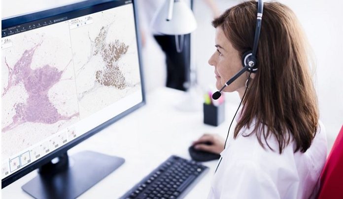
International medical imaging IT and cybersecurity company Sectra has deployed its software for medical imaging at Leeds Teaching Hospitals. It is the first of an initial six trusts in the Northern Pathology Imaging Co-operative (NPIC) to go live, paving the way for one of the most sophisticated interconnected pathology initiatives in the world. The technology from Sectra will underpin the regional program that is digitizing, connecting and applying artificial intelligence to pathology services in the North of England.

Leeds Teaching Hospitals went live with Sectra’s solution for digital pathology and Vendor Neutral Archive (VNA) in October. This will allow pathology images to be interrogated by professionals electronically from a range of devices-anywhere and at any time. With many pathology departments across the country still reliant on microscopes and glass slides, the new system is allowing pathologists to work more quickly, gain easier access to opinions from colleagues and manage rising demand.

Furthermore, the joint VNA will allow the trust to pool imaging and build a platform for artificial intelligence that is crucial to improving diagnoses for cancer and other illnesses.

“Leeds is the first of our six sites to go fully digital. Collectively, we are modernizing our pathology services to become among the most advanced and interconnected anywhere in the world, and we hope to share our experience to help others across the NHS and beyond,” says Dr. Darren Treanor, NPIC’s director, and a practicing pathologist at Leeds Teaching Hospitals NHS Trust.

He continues: “The days of using glass slides and paper notes to determine and communicate a patient’s diagnosis are numbered. As we move to digital ways of working, we can improve quality and create a more structured digital workflow.”

Greater benefits are expected as more hospitals in the NPIC go live with the system later in the year. As announced in 2018, the program is part of a partnership between industry, the NHS and academia, funded by UK Research and Innovation and industry partners to connect pathology services across the region using technology. This will lead to the full digitization of NHS laboratories covering a population of three million people, allowing hospitals to pool resources, balance workload, and enable easier access to specialists across the region whose expertise may be quickly needed to make a clinical diagnosis.

“Digital pathology is about far more than replacing microscopes with computers. It’s about fundamentally changing how pathology services can be configured across regions and across the country, so that patients can receive faster diagnoses, services can become more intelligent, and the NHS can make best use of its valuable pathologists. NPIC is at the forefront of this transformation,” says Jane Rendall, UK Managing Director for Sectra.

About NPIC

Northern Pathology Imaging Co-operative, NPIC (Project no. 104687) is one of five centers supported by a ยฃ50m investment from the Data to Early Diagnosis and Precision Medicine strand of the government’s Industrial Strategy Challenge Fund, managed and delivered by UK Research and Innovation (UKRI), announced in 2018.

NPIC is a unique collaboration between NHS, academic and industry partners. NPIC is deploying digital pathology across hospitals in the North of England and will develop artificial intelligence tools to help diagnose cancer and other diseases.

About Sectra

Sectra assists hospitals throughout the world to enhance the efficiency of care, and authorities and defense forces in Europe to protect society’s most sensitive information. Thereby, Sectra contributes to a healthier and safer society. The company was founded in 1978, has its head office in Linkรถping, Sweden, with direct sales in 19 countries, and operates through partners worldwide. Sales in the 2019/2020 fiscal year totaled SEK 1,661 million.

Sony Medical Imaging Platform NUCLeUS new functionalities support remote patient observation

 Sony Medical Imaging Platform NUCLeUS new functionalities support remote patient observation

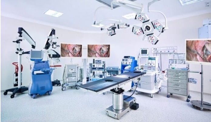
Sony has announced an update to its vendor-neutral medical imaging platform NUCLeUS. This version built in direct response to the pandemic, introduces remote patient observation with new recording functionalities for use in the operating room (OR), Intensive Care Units (ICU), endoscopy suites, interventional rooms or anywhere else in the hospital.

Developed in consultation with leading surgeons and with vendor neutrality in mind, NUCLeUS guides clinical staff through the planning, recording and sharing of any type of video, still images and other patient-related data. Seamlessly linking devices, applications, video and most importantly, people, NUCLeUS focuses on hospital staff requirements and use cases, adding value to imaging workflows.

As of today, NUCLeUS has been enhanced with new functionalities and features, including improved bi-directional telestration allowing multiple remote users to simultaneously annotate, draw or highlight areas of interest in a live stream video. This can be shared by viewers in all locations to discuss as a group in real time, ideally suited to socially distanced set-ups. In addition, new recording functionalities have also been added as a core use for Ear, Nose and Throat, Urology, Obstetrics and Ophthalmic as well as Outpatient/Ambulatory Surgical Centers (ASC) and private practice uses, including plastic surgery, colorectal and more.

โ€œSony is committed to developing NUCLeUS to suit the needs of the patient and medical staff at all times. The remote patient observation is a good example of this as it was brought in to help hospitals manage day-to-day requirements through the Covid-19 pandemic,โ€ explained Ludger Philippsen, head of Healthcare at Sony Professional Solutions Europe. โ€œIn the wake of the global pandemic we have helped hospitals and healthcare providers reinvent their workflows, providing medical staff with the tools they need to continue delivering excellent patient care.โ€

New functionalities of NUCLeUS include:

  • Mosaic Video Wall, presenting video streams from image sources in multiple ORs and ICUs simultaneously on a single display, thus providing a situational overview of activity in a tiled or mosaic format.
  • An iPad Streaming function, allowing clinical staff to access images from any modality via an iPad in virtual real time within the OR, so medical staff can follow the intervention on their handheld device.
  • High quality 4K conversion, allowing any HD resolution video content to be converted 4K using advanced resolution-augmentation algorithms superior to conventional upscaling, giving a crisp ultra-high resolution view of converted video footage.
  • Customizable Expanded Patient Distraction โ€“ helping to reduce patient anxiety through music tracks and video imagery that can be played in the OR to create a more relaxing and comfortable atmosphere.
  • Patient Time-Out Functionality, featuring checklists that simplify time out of safety standards at the start, during and end of an operation.
  • Enhanced Printing capabilities, allowing hard copies of still images captured by NUCLeUS to be created inside the OR using an optional UP-DR80MD A4 digital colour printer. The Auto Print function also extends CMS (Content Management System) print functionality to collect a preconfigured number of stills, printing them automatically when this number of images has been captured.
  • Full compatibility with the latest Sony PTZ and fixed cameras including HD and 4K models

As healthcare embraces digitalization, 2020 has been a game changer for our industry.

Proven Technology VCS and Medical Physicians Launch Decentralized Health Data Marketplace Powered by Blockchain and AI Technology

The IKIGAI Network is pioneering the OpenHealth movement by building the worldโ€™s first global decentralized healthcare ecosystem built on principles of open data and open source development. This new information infrastructure for healthcare will allow anyone to synchronize health data with a network of physicians, individuals, entrepreneurs, and institutions. With a more accessible, efficient, and smarter health information management system not siloed by any one institution, IKIGAI will enable a thriving ecosystem for patients and clinicians around the world.

Unlocking personal health data allows patients to benefit from their health data in new, innovative ways and provides timely access to data for better clinical outcomes. Artificial intelligence will be leveraged to Blockchain consensually run on individualsโ€™ health data to provide hyper-personalized, proactive healthcare recommendations and unlock large real-time health datasets for medical researchers.

Verida is a software company that leads the development of self-sovereign data technologies for individuals and enterprises, founded by Chris Were, former CEO and co-founder of Australian technology company Community Data Solutions. With 20+ years of experience in software solutions, Chris started Verida as a research project in 2018, with the mission of helping people own and control their data. โ€œWe created an open-source library that enables decentralized applications to be built where users can own, control, and store their data using private encryption keys,โ€ says Were.

Recommended AI News: Innovaccerโ€™s Member Engagement Solution Now Available on Microsoft Azure

CareProtocol is an AI health start-up founded by two physicians, Dr. Roger Ng and Dr. Ray Ng, who hold 20+ years of cumulative practice experience, and two technology entrepreneurs and proven investors, Marc van der Chijs and Sean Clark. Dr. Ray Ng believes โ€œthe joint partnership is a natural fit for these two organizations to join forces to launch IKIGAI, as both teams share the goal of improving the state of healthcare, empowering both physicians and patients.โ€

According to Sean Clark, CEO of IKIGAI, โ€œthe healthcare industry is the largest economic sector in developed economies, representing nearly $4 trillion in the U.S. alone. With the emergence of COVID-19 driving growth in telehealth services, the rise of Blockchain decentralized currencies, and a growing mistrust of centralized systems amidst a global pandemic, the time is right for decentralized healthcare.โ€

Members of the IKIGAI Network will be able to access a diverse range of OpenHealth services built on top of an open-source framework delivering the worldโ€™s first decentralized App Store (dApp Store) for healthcare. dApps available at launch will include HealthOS by Care Protocol, Decentralized EMR and COVID19 Credentials by Verida, and the IKIGAI Vault, which enriches the health experience by allowing patients to own, control and consensually share their personal health data with experts around the world.

Connect Health introduces new COVID-19 musculoskeletal risk assessment tool

Connect Health introduces new COVID-19 musculoskeletal risk assessment tool

CHART (COVID-19 Connect Health Assessment of Risk Tool) introduced to support MSK clinicians in making the right decisions for managing patient care whilst services are restricted

A new risk assessment tool which supports MSK clinicians in making difficult decisions in weighing up COVID-19 risk with clinical need, has been rolled out nationwide and published in the BMJ Open Sport & Exercise Medicine. The journal, which has a strong MSK readership worldwide, published C-CHART, detailing how the โ€˜tool can serve to improve the consistency of clinical decision-making during restitution of face-to-face MSK services in Englandโ€™.

As the pandemic caused clinicians to switch from face-to-face musculoskeletal (MSK) care to providing telephone or video consultations almost overnight, Connect Health, the UKโ€™s leading independent community provider of musculoskeletal services (including physiotherapy, orthopedics, pain, rheumatology, and occupational health services), initially designed C-CHART to guide their own MSK clinicians to balance the health needs of patients whilst prioritizing patient safety during the protracted Covid-19 outbreak.

Having trialed it for a number of months across the UK and using the latest evidence from UK studies, Connect Health has now made the tool available to all clinicians, expertly reducing subjectivity in assessment of risk and improving the consistency of clinical decision-making during restitution of face to face MSK services.

As waiting lists for planned care rise – reports suggest from 4m to 10m by the end of this year, NHS England has recently recommended prioritization of more urgent conditions, including those people whose condition has deteriorated and those waiting the longest, as part of a phased return to pre-COVID-19 service provision. Routine GP appointments are slowly getting back to near normal levels, and the pandemic has led to significant changes in how NHS services are delivered and the vital need for infection control.

Clinicians now need to assess an individualโ€™s risk factors for complications from COVID-19 alongside their clinical priority to inform a shared-decision making discussion about appropriate face to face care delivery.

There are two parts to the tool; the first is the individual patient COVID risk assessment and the second is the clinical prioritization, enabling the clinician to answer questions related to patient risk and the increased risk to the clinicians of a face-to-face appointment. The resulting overall risk score informs a shared decision-making discussion with the patient about the balance of risks and benefits of in-person care and aims to reduce subjectivity.

Scientists use new approach to chart the spread of drug-resistant hospital bacteria

Colorectal cancer on an upswing in South India

Scientists have used genome sequencing to reveal the extent to which a drug-resistant gastrointestinal bacterium can spread within a hospital, highlighting the challenge hospitals face in controlling infections.

Enterococcus faecium is a bacterium commonly found in the gastrointestinal tract, where it usually resides without causing the host problems. However, in immunocompromised patients, it can lead to potentially life-threatening infection.

Over the last three decades, strains have emerged that are resistant to frontline antibiotics including ampicillin and vancomycin, limiting treatment options – and particularly worrying, these strains are often those found in hospital-acquired E. faecium infections.

A team of scientists at the University of Cambridge and the London School of Hygiene and Tropical Medicine has pioneered an approach combining epidemiological and genomic information to chart the spread of bacteria within healthcare settings. This has helped hospitals identify sources of infection and inform infection control measures.

In a study published today in Nature Microbiology, the team has applied this technique to the spread of drug-resistant E. faecium in a hospital setting.

“We’ve known for over two decades that patients in hospital can catch and spread drug-resistant E. faecium. Preventing its spread requires us to understand where the bacteria lives – its ‘reservoirs’ – and how it is transmitted.

The team followed 149 hematology patients admitted to Addenbrooke’s Hospital, Cambridge University Hospitals NHS Foundation Trust, over a six-month period. They took stool samples from the patients and swabs from the hospital environment and cultured them for E. faecium.

Genomic analysis of the bacteria was much more effective at identifying hospital-acquired E. faecium: out of 101 patients who could be followed up, genomic analysis identified that two thirds of patients acquired E. faecium, compared to less than half using culture methods alone.

Just under half (48%) of the swabs taken from the hospital environment were positive for vancomycin-resistant E. faecium. This included 36% of medical devices, 76% of non-touch areas such as air vents, 41% of bed spaces and 68% of communal bathrooms tested.

The researchers showed that even deep cleaning could not eradicate the bacteria. The hospital undertook deep cleaning on one ward over a three-day period during the study, when patients were moved elsewhere; however, when the team sampled locations prior to patients returning to the ward, they found that 9% of samples still tested positive for the bacteria. Within three days of patients returning to the ward, around half of the sampled sites tested positive.

Three-quarters (74%) of the patients (111/149) were carriers of the A1 clade – a multi-drug resistant strain of E. faecium commonly seen in hospitals that is resistant to the antibiotic ampicillin and which frequently acquires resistance to vancomycin. Of these 111 patients, 67 had strong epidemiological and genomic links with at least one other patient and/or their direct environment.

“The fact that these cases were all linked to another patient or their environment suggests strongly that they had picked up the multi-drug resistant bacteria while in the hospital,” said Dr Francesc Coll from the London School of Hygiene and Tropical Medicine, joint first author.

Further genomic analysis showed that within this multi-drug resistant strain were several subtypes (defined by how genetically-similar they were). However, it was not uncommon for a patient to be carrying more than one subtype, which – without detailed genomic analysis – could confound attempts to identify the route of transmission of an infection.

Notably, despite the circulation of as many as 115 subtypes, 28% of E. faecium acquisitions were caused by just two superspreading subtypes. The authors found no evidence of resistance or tolerance to common disinfectants to explain the success of these subtypes.

Six study patients contracted an ‘invasive infection’, meaning that they had been carrying E. faecium asymptomatically in their gut, but subsequently developed a symptomatic infection. Comparing the genomes of the infecting and gut strains the authors determined that invasive E. faecium infections originated from the patients’ own gut.

“Our study builds on previous observations that drug-resistant strains of E. faecium can persist in the hospital environment despite standard cleaning – we were still surprised to find how short-lasting was the effect of deep cleaning,” added Dr Gouliouris.

“We found high levels of hospital-adapted E. faecium despite the use of cleaning products and procedures that have proven effective against the bug. It highlights how challenging it can be to tackle outbreaks in hospitals.”

Senior author Professor Sharon Peacock from the Department of Medicine at the University of Cambridge added: “The high rates of infection with drug-resistant E. faecium in specific vulnerable patient groups and its ability to evade cleaning measures pose an important challenge to infection control.”

“Patient screening, adequate provision of isolation and ensuite toilet facilities, improved and more frequent cleaning procedures, and stricter health-care worker hygiene practices will all be needed to curtail this global epidemic.

“But this is also a sign of how urgently we need to tackle inappropriate use of antibiotics worldwide, which is widely recognized as posing a catastrophic threat to our health and our ability to control infections.”


 

Teleradiologists accuracy diagnosing COVID-19 underscores remote readings importance during emergencies

Teleradiologists with varying levels of experience are highly accurate at diagnosing COVID-19 on chest CTs, according to a new multi-center study. It should give hospitals more confidence to deploy outsourced emergency reading during the pandemic.

Thatโ€™s according to a large group of radiologists out of France, who compared remote reads performed by on-call experts to those completed by senior imaging physicians.

In more than 500 adults with confirmed COVID-19, teleradsโ€™ diagnoses were โ€œhighlyโ€ consistent with patientsโ€™ reverse transcription-polymerase chain-reaction (RT-PCR) testing results, the authors wrote Oct. 29 in European Radiology.

Senior rads also agreed with their remote peers in most instances, first author Hubert Nivet, with Imadis Teleradiology in Lyon, France, and colleagues noted.

While a majority of radiology societies do not recommend using CT as a first-line tool to screen patients for the novel virus, the authors believe their findings may serve as a template of sorts for emergency situations.

โ€œOur results illustrate the high diagnostic accuracy โ€ฆ of teleradiologists with various degrees of experience, in settings with different levels of prevalence, as well as an excellent interobserver agreement for chest CT,โ€ Nivet et al. added. โ€œThus, this kind of structured outsourced teleradiology model could [pair] high-quality structured and standardized reports with report turnaround time[s] meeting the requirements of emergency medicine during the pandemic.โ€

For their research, the group prospectively enrolled 513 patients who received both a chest CT and RT-PCR testing at one of 15 hospitals. Of that population, 244 (47.6%) came back with a lab test positive for COVID-19.

All scans were immediately read by on-call teleradiologists, which consisted of 106 senior rads and 45 junior radiologists, and then blind reviewed by a team of 15 senior radiologists. The study took place between March 13 and April 14.

Overall agreement between initial emergency interpretations and secondary reads was excellent, the authors noted (weighted kappa=0.87). Interpretations also correlated with lab testing results.

The study was limited by a number of factors, including variable COVID-19 prevalence rates between hospitals, the authors noted.

โ€œChest CT demonstrated high diagnostic accuracy with strong interobserver agreement between on-call teleradiologists with varying degrees of experience and senior radiologists over the study period,โ€ Nivet and colleagues concluded.

Philips expands its home care portfolio for COPD patients with first-of-its-kind non-invasive ventilator

Philips expands its home care portfolio for COPD patients with first-of-its-kind non-invasive ventilator

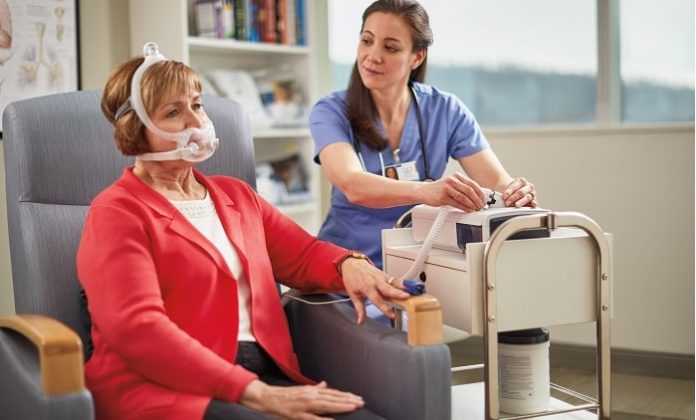
Royal Philips, a global leader in health technology, announced the launch of Philips Ventilator BiPAP A40 EFL. With the introduction of this non-invasive ventilator, Philips extends its homecare solutions with a new ventilation therapy feature for chronic obstructive pulmonary disease (COPD) patients to breathe easier. Now, pulmonologists can identify COPD patients with expiratory flow limitation (EFL) and treat them with targeted therapy to reduce symptoms and increase their comfort while sleeping. The BiPAP A40 EFL ventilator continuously and optimally adjusts pressure based on patient needs.

BiPAP A40 EFL is the first and only non-invasive ventilator that allows health care professionals to automatically screen for and detect EFL, then provide optimal homecare therapy to dynamically and automatically abolish EFL [1]. This helps to reduce the patientโ€™s work of breathing. Built with Philips proprietary and clinically validated ExpiraFlow technology, BiPAP A40 EFL is designed to connect across the care pathway โ€“ from diagnostic work to point of care therapy โ€“ to enable informed clinical decisions and optimize ventilation therapy, even remotely.

More than 50 percent of COPD patients experience EFL โ€“ limited exhalation of breath from the lungs โ€“ which occurs in the lower airways when patients are breathing quietly [2]. EFL causes hyperinflation, or breathing at increased lung volumes. COPD patients with EFL are more likely to be hospitalized more often and have increased mortality rates, however EFL is difficult to detect and often undertreated, despite its prevalence. Philips unique ExpiraFlow technology automatically detects EFL more accurately than any alternate methods [3] to enable the more effective treatment of patient in the home and help avoid hospital readmissions [2].


โ€œExpiraFlow Technology represents a shift in the paradigm of ventilator COPD management toward more personalized therapy, which automatically optimizes ventilation to the individual needs of the patient,โ€ said Peter Calverley, Professor of Respiratory Medicine, School of Aging and Chronic Disease at the University of Liverpool. โ€œBy monitoring the presence of EFL on a breath-by-breath basis, the A40 EFL system can automatically adjust therapy pressures to ensure efficient lung emptying and better gas exchange. This new focus allows us to consider individual differences in lung mechanics and gas exchange when managing complex respiratory patients.โ€

โ€œEFL often goes undetected, meaning patients donโ€™t receive the care they need to improve their disease,โ€ said Eli Diacopoulos, Respiratory Care Business Leader at Philips. โ€œAt Philips, weโ€™re committed to identifying these gaps and meeting the challenges that COPD patients face every day. BiPAP A40 EFL aims to revolutionize COPD care solutions. Clinicians can now detect EFL in hypercapnic COPD patients at the point of care, ensure personalized patient treatment at home and monitor care remotely.โ€

The BiPAP A40 EFL leverages Philips leading connected solution platform to streamline diagnostic work through integration to Philips Alice sleep lab and home diagnostic systems. When prescribed and used

in the home, the BiPAP A40 EFL connects to Philips Care Orchestrator cloud-based care management system. By making it easier to analyze and share information [4], this connectivity enables providers to make faster, more informed clinical decisions, and identify and prioritize patients who are in need of therapy intervention to better manage chronic respiratory patient care from hospital to home.

Translate ยป