

















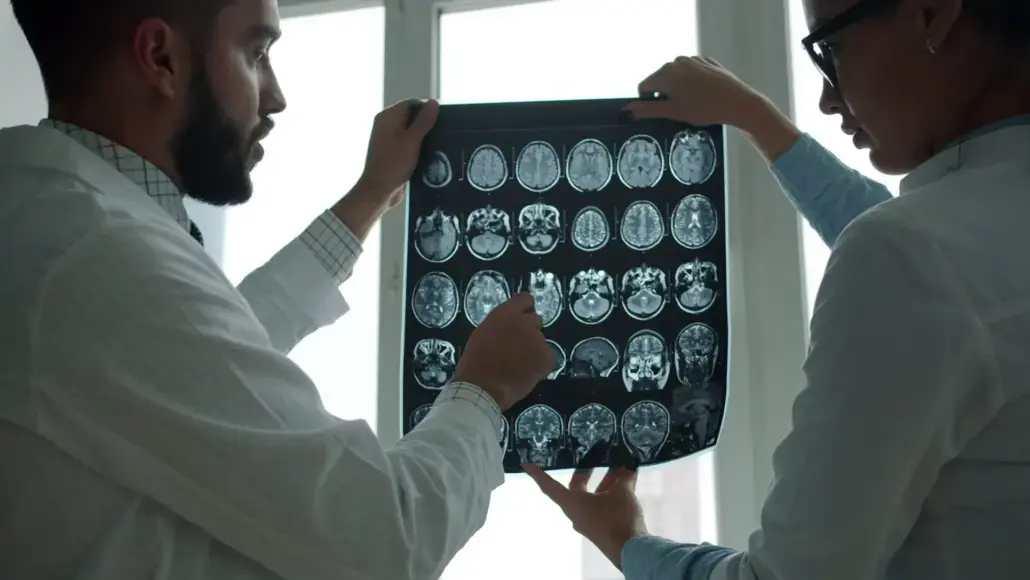

A brain scan can look unremarkable while the changes researchers care about are already underway. That creates a difficult gap in neurodegenerative disease: the first noticeable memory problem isn’t necessarily the first sign of damage, and a reassuring visual impression doesn’t settle what has changed beneath it.
Detecting microscopic CNS changes before symptoms appear raises a question about what imaging actually measures. In Alzheimer’s disease, researchers want to understand changes in the central nervous system (CNS) before everyday difficulties make them obvious. Yet structural MRI doesn’t show individual neurons, and brain atrophy isn’t always apparent from looking at a scan.
The growing interest in deep learning sits within that tension. Earlier detection depends on distinguishing meaningful structural change from ordinary variation, without treating every small difference as evidence of disease.
The Short Answer: Brain Structure Shifts Before Memory Does
Microscopic CNS changes can be detected before symptoms appear, with an important limit: neuroimaging can identify structural patterns and trajectories, but it doesn’t directly count microscopic neuronal losses. Deep-learning analysis extends what researchers can extract from scans without removing the need for clinical interpretation.
In preclinical Alzheimer’s, underlying disease processes can develop before obvious cognitive symptoms. The familiar phrase “early symptoms” therefore describes when difficulties become apparent, not necessarily when the biological process began. By the time cognitive symptoms appear in conditions such as Alzheimer’s disease, millions of neurons have already been lost.
Neuroimaging’s technical evolution shifts the question from “Does this scan look abnormal?” to “What changed, where, and by how much?” In clinical trials, that means identifying participants whose disease biology and stage match the research question, then selecting measurements that can test a treatment’s effect.
Why Symptoms Are a Late Signal of Neurodegeneration
Neurodegeneration and noticeable impairment don’t follow identical timelines. Symptoms aren’t a direct measurement of brain tissue. Mild cognitive impairment describes a clinical state, not a precise quantity of neuronal loss. Symptom recognition and structural measurement answer related but different questions.
Everyday Forgetfulness Versus Meaningful Early Change
Repeating questions, misplacing belongings, and struggling to find words commonly feature in descriptions of early Alzheimer’s symptoms. However, occasional lapses also occur with ordinary ageing, stress, and distraction. A single forgotten name doesn’t distinguish those explanations from neurodegeneration.
The more informative distinction is the pattern: whether difficulties persist, progress, and interfere with familiar activities. Short-term memory loss carries a different significance when someone repeatedly loses track of recent conversations than when an isolated detail slips their mind.
Where Imaging Fits on the Alzheimer’s Stage Timeline
The Global Deterioration Scale offers a seven-stage framework spanning no apparent cognitive decline through severe impairment. It describes clinical progression; it isn’t a timetable that assigns a fixed duration or imaging signature to each stage.
Biomarkers approach the problem from another direction. The biomarker research describes efforts to identify underlying Alzheimer’s-related changes, including changes that precede symptoms.
Structural imaging belongs within that broader evidence picture. It measures anatomy rather than memory performance, giving researchers information that symptom reports alone can’t provide. Importantly, an anatomical difference isn’t automatically specific to Alzheimer’s, so interpretation requires more than a volume measurement.
From the Radiologist’s Eye to Deep-Learning Analysis
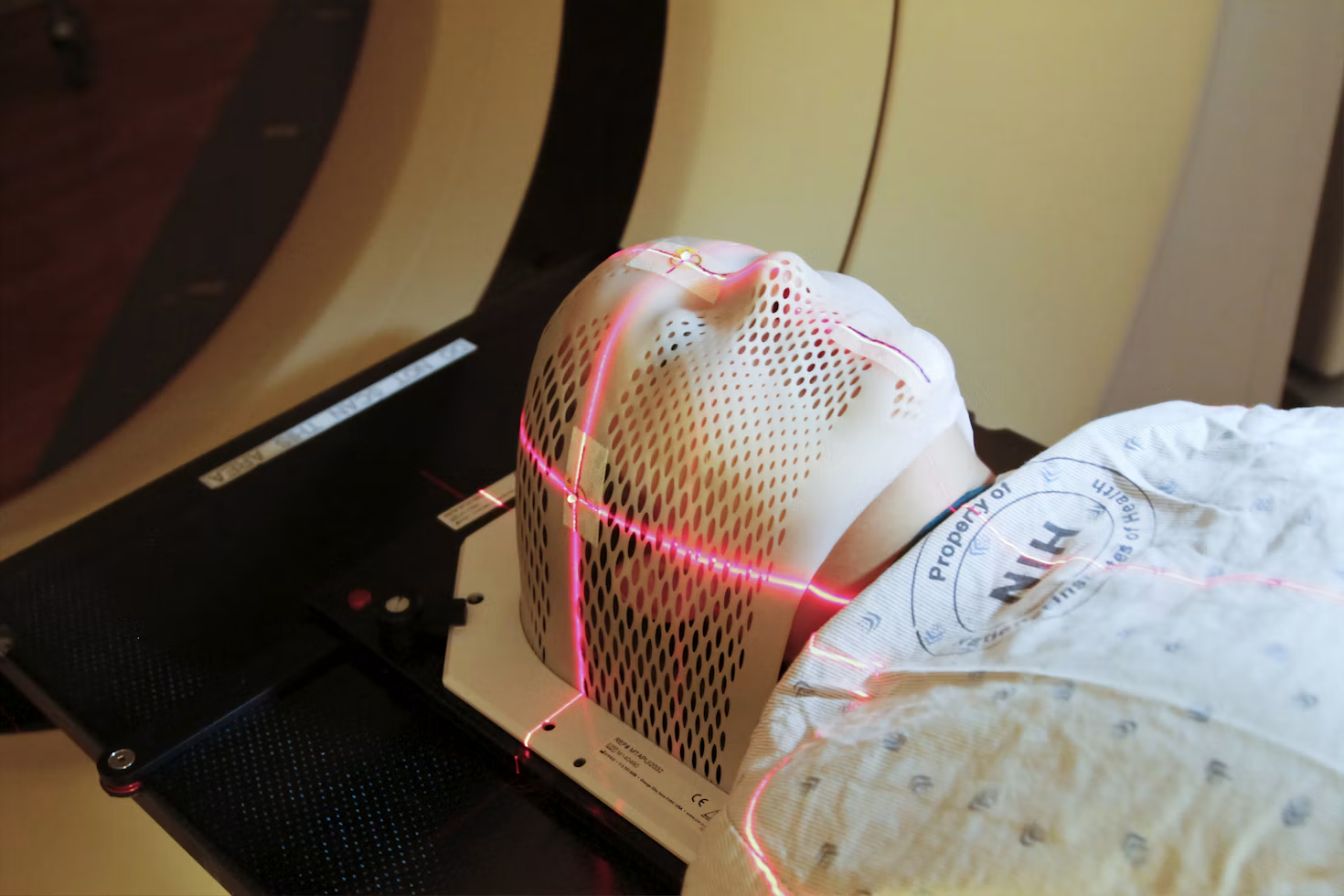
Within that shift from looking to measuring, neuroimaging data analytics converts MRI images into measurements that researchers can compare over time. Volumetric MRI adds numerical descriptions of brain structures to the visual assessment, rather than making that assessment redundant.
What a Qualitative Read Can Miss in Subtle Atrophy
A radiologist assesses anatomy, image quality, and patterns of abnormality. Visual rating scales can organise judgments about atrophy, but they still ask a reader to place continuous anatomical variation into categories.
Subtle change presents a different problem from obvious tissue loss. Diffuse or symmetric shrinkage might not produce a conspicuous contrast, and two scans viewed side by side don’t directly quantify their difference. Reader judgment also introduces variation, particularly around category boundaries.
Quantitative analysis instead estimates the volume of defined structures, including the hippocampus and entorhinal cortex. Repeated measurements can describe a trajectory that a single scan cannot establish.
The comparison still needs context. A small reported volume difference could reflect tissue change, acquisition differences, or an imperfect boundary estimate. A number isn’t meaningful simply because software produced it.
How Algorithms Detect Sub-Pixel Shifts in Brain Volume
A voxel is a three-dimensional sampling element in an MRI image. “Sub-pixel atrophy” shouldn’t imply that MRI resolves microscopic cells.
In a three-dimensional scan, the more appropriate concept is estimating changes smaller than a voxel’s dimensions from information distributed across many voxels.
Deep-learning models, including convolutional neural networks, learn patterns from training scans to help identify tissue and anatomical boundaries. Registration aligns serial images, while segmentation assigns image regions to structures whose volumes can be estimated. These tasks turn visual differences into measurable quantities.
Such estimates aren’t direct observations of individual neurons, and deep learning doesn’t automatically guarantee reliable sub-voxel measurement.
Image harmonisation addresses differences between scanners, acquisition settings, and sites. Validation must then establish whether the measured changes are repeatable and relevant to clinical outcomes.
Algorithms complement radiologists by supplying measurements; radiologists and research teams remain responsible for interpreting their significance.
What Earlier Detection Changes for Drug Trials and Patients
An earlier signal matters when it changes a decision. However, detecting a smaller anatomical difference isn’t, by itself, evidence that treatment will improve someone’s life.
Stratifying Participants Before Irreversible Decline
Patient stratification groups participants by characteristics relevant to a trial. Structural measurements can contribute information about the extent and pattern of tissue loss, alongside cognitive assessments and biomarkers such as amyloid.
This helps researchers distinguish an early-disease study population from one with more established damage. It also avoids treating everyone with a similar memory complaint as biologically interchangeable.
Disease-modifying therapies require trials designed around the disease process they target, not symptoms alone.
Atrophy measurements can also serve as trial endpoints, but their role needs careful definition. A change in brain volume doesn’t automatically equal a change in clinical benefit.
More precise measurements could support more efficient studies if validation demonstrates lower measurement variability and a meaningful treatment signal. Smaller cohorts or shorter studies aren’t guaranteed consequences of adding an algorithm.
Can Catching Changes Early Reverse Dementia?
Earlier detection isn’t reversal. Identifying structural loss sooner doesn’t restore lost neurons, and detecting an imaging change doesn’t establish that a treatment can undo it.
For established neurodegenerative disease, slowing progression and reversing damage are different goals.
Cognitive symptoms also aren’t synonymous with neurodegeneration. Clinical assessment distinguishes degenerative disease from other contributors, including vitamin deficiencies, thyroid problems, and medication effects.
Improvement in a treatable contributor shouldn’t be confused with reversal of Alzheimer’s-related tissue loss.
For individuals, an earlier, well-supported finding can inform monitoring, planning, and trial eligibility. Its value depends on the reliability of the evidence and the decisions available, not simply on how early a scan produces a flag.
Reading the Brain Before It Tells Us Something Is Wrong
The gap between biological change and noticeable neurodegeneration creates an opportunity to study intervention earlier. Trial stratification is a concrete use of that opportunity: matching participants to disease stage and measuring change consistently.
For research and clinical practice, the meaningful advance isn’t merely seeing more. It’s knowing which measured differences justify a different decision.