Close
Digital Health & Ai Innovation summit 2026
Medical Taiwan 2026

Caretaker Medical Adds ECG Patch to the Caretaker Wireless Continuous Blood Pressure & Patient Monitoring Platform

Caretaker Medical Adds ECG Patch to the Caretaker Wireless Continuous Blood Pressure & Patient Monitoring Platform

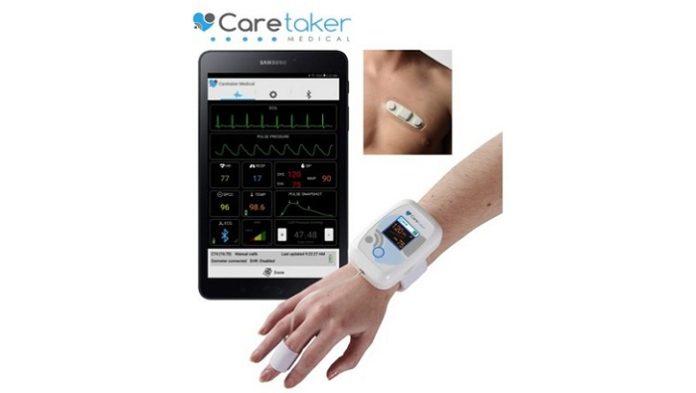
Caretaker Medical, a leader in clinical-grade wireless patient monitoring and maker of the Caretakerยฎ wearable patient monitor that utilizes a simple Finger Cuff to measure Real-time โ€œBeat by Beatโ€ Continuous Blood Pressure, Respiration Rate, and other hemodynamic parameters, has incorporated VivaLNKโ€™s FDA-Cleared ECG Patch to their wireless patient monitoring platform.

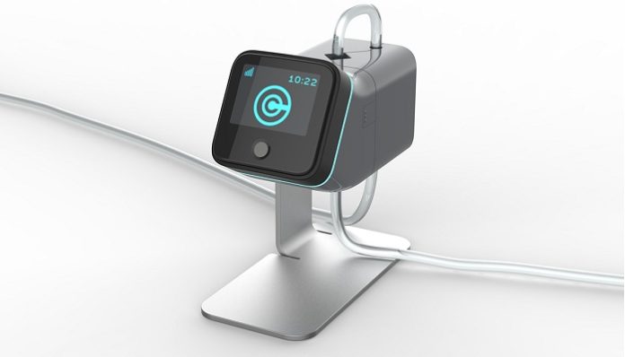
Caretaker Continuous Blood Pressure & Wireless Vital Signs Monitor with ECG Patch

The Caretakerยฎ patient monitor provides clinicians with continuous, uninterrupted Blood Pressure and Vital Signs data across the full continuum of care in a completely wire-free form factor that maximizes patient mobility and provides a comprehensive view of hemodynamic health. Clinicians can remotely view patient data from virtually anywhere using the Caretaker App, the CaretakerCloud Remote Monitoring Portal, or via data integration with other monitoring platforms.

โ€œOur patented Pulse Decomposition Analysis method of non-invasive hemodynamic measurements like continuous beat-by-beat Blood Pressure provides clinicians with a view of the patientโ€™s mechanical heart function, and VivaLNKโ€™s reusable ECG Patch reveals the status of the patientโ€™s electrical heart function,โ€ said Jeff Pompeo, Caretaker Medicalโ€™s President & CEO. โ€œCombined, the Caretaker monitoring platform now provides a seamless view of both electrical and mechanical heart health, including waveforms, in a completely wire-free, wearable form factor that doesnโ€™t inhibit patient mobility.โ€

Both the Caretaker wireless monitor and the VivaLNK ECG Patch are FDA-Cleared and CE-Mark certified and available for purchase from Caretaker Medical and its distributors. For more information please visit www.CaretakerMedical.net

About Caretaker Medical
Caretaker Medical is a wireless medical device firm focused on developing affordable, innovative, patient monitoring solutions that improve outcomes, increase clinician productivity, and maximize patient mobility. The wearable Caretaker4ยฎ monitor enables uninterrupted wire-free and electrode-free vital signs monitoring throughout the full continuum of care, from clinic and hospital, to patient transport, and home monitoring. Using a low-pressure finger cuff, Caretakerโ€™s patented Pulse Decomposition Analysis technology non-invasively measures continuous Beat-by-Beat Blood Pressure, and Vital Signs for remote display on the Caretakerยฎ App and other Remote Data Displays.

Cynerio Introduces Virtual Segmentation to Healthcare IoT Cybersecurity

Cynerio Introduces Virtual Segmentation to Healthcare IoT Cybersecurity

Cynerio, a leading provider of healthcare IoT cybersecurity solutions, announced the addition of the virtual segmentation capability to their platform. Safe and effective healthcare IoT security projects can take over a year to execute due to a lack of visibility into network topology and sensitive device operations.

Building effective cybersecurity policy requires thorough analysis and the ability to contextualize and prioritize devices according to impact. The Cynerio platformโ€™s new virtual segmentation capability automatically delivers safe and effective policies in a matter of weeks by customizing segmentation policy for every device type, limiting the attack surface, and ensuring clinical services remain intact.

The virtual segmentation capability profiles device communications and generates East-West and North-South segmentation policies, allowing IT security teams to test the policies and monitor for violations before pushing it to live clinical network environments. The AI-based technology groups devices and segments them according to network behavior while risk is prioritized based on device criticality and medical impact. The platform then monitors the network to ensure all traffic is compliant with policy, and provides tools to refine it, sending automatic updates when policy is safe to push to firewall and NAC.

“The challenge of achieving safe and effective segmentation is what inspired us to develop the virtual segmentation capability for our customers,” states Leon Lerman, CEO and co-founder of Cynerio. “Our top priority is making healthcare IoT security projects as easy and painless as possible by helping hospitals safely navigate the risks of immediately enforcing segmentation policies in clinical environments. Cynerio already offers seamless integration with top-tier cybersecurity tools like Cisco, Check Point, Forescout, Palo Alto, and more. Adding virtual segmentation to our platform saves hospitals years of time and resources spent on IoT security projects and provides policies that can be easily enforced with confidence.”

Cynerio is a premier sponsor of the HIMSS 2020 Cybersecurity Forum where CEO and co-founder, Leon Lerman, will present โ€œTaking a Medical-First Approach to Solving Tomorrow’s IoMT Challengesโ€ on March 9.

About Cynerio

Cynerio is the world’s premier medical-first IoT cybersecurity solution. We view cybersecurity as a standard part of patient care and provide healthcare delivery organizations with the insight and tools they need to secure clinical ecosystems and achieve long-term, scalable threat remediation without disrupting operations or the delivery of care.

The Benefits Of Online Scheduling For Healthcare Practices

The Benefits Of Online Scheduling For Healthcare Practices

In an increasingly technology-driven world, healthcare providers are now expected to provide easy and convenient facilities, as well as high-quality care. Online scheduling is a software tool that enables patients to book or request appointments with only a few simple clicks from any internet-connected device. It is one of the most popular and simplest technologies used by forward-thinking healthcare providers, including hospitals. Using online scheduling can bring many benefits to practices who use it.

Increase In New Patients

People are becoming less inclined to make phone calls. Instead, patients prefer using a website or an app to book an appointment, rather than call the practice. In some cases, patients may feel embarrassed to discuss their problem over the phone. As a result, new patients looking for a new doctor or specialist tend to be more attracted to those who provide online scheduling. This gives them the freedom to book appointments themselves, without having to ring up someone they donโ€™t know.

Reduces Empty Appointment Slots

By using online scheduling, healthcare practices are much more likely to fill empty appointment slots. For the practice this is a great benefit, as empty appointment slots result in lost revenue. For patients, it enables them to book same-day or next-day appointments quickly, whether this be late in the evening or first thing in the morning. They can quickly check a practiceโ€™s availability and book an appointment that suits them.

24-Hour Availability

Previously, patients could only book appointments during a clinicโ€™s opening hours. Yet with online scheduling they are now able to log in to the clinicโ€™s website at any time of day, on any day of the week. They are able to check a doctorโ€™s availability and make an appointment at a time that suits them and allows them to plan their schedules more efficiently around their visit.

More Effective Information Management

Online systems make managing patient information much easier and more efficient. Patient health records management is included in some online appointment scheduling systems. โ€œA practice can create a single point from which patient information can be saved, updated and managed,โ€ says Robert Monroe, a digital technology blogger at DraftBeyond and Research Papers UK. โ€œCrucially, this allows for more efficient analysis and better managements of a patientโ€™s case file.โ€

Increased Opportunities for E-marketing

One of the benefits of online scheduling is that it can include the phone numbers and email addresses of patients. As a result, promotional drives (such as blood test packages, free check-ups or healthcare packages) can be quickly circulated to patients, using the information already acquired via the online scheduling system. Patients are more likely to keep these details up-to-date when using the software, which means that there is a greater chance it will definitely reach the patient. This can lead to more visits being made by existing patients or the registration of new patients through references.

Easier Method of Payment Online

Patients who book appointments online are less likely to miss or even avoid their appointments, as all services offered will have an associated payment related to them. โ€œPatients tend to feel comfortable making the payments online because the online scheduling systems are safe and the information stored is kept securely,โ€ says Katherine Jackson, a healthcare writer at Lucky Assignments and Gum Essays. โ€œYou can also encourage patients to use the scheduling services more often by making offers such discounts at set times or offering a free consultation after the first visit.โ€

Reduction in Administrative Work

Front office staff can find it challenging to provide quality service to patients if they are trying to multi-task billing, administrative tasks and answering phone calls. This can lead to long hold times and even curt customer service, which can result in unhappy patients. With online scheduling, staff are able to better spend their time providing a positive experience for patients who are physically in the practice, as well as freeing up time to complete more complex work.

Using online appointment scheduling systems offers wide-ranging benefits for both patients and healthcare providers. It enables practices to update and improve their practiceโ€™s services and allows them to devote more time to offering quality patient care. Online scheduling offers patients consistency and predictability.

Explore Carestreams OnSight 3D Extremity System at ECR

Explore Carestreams OnSight 3D Extremity System at ECR

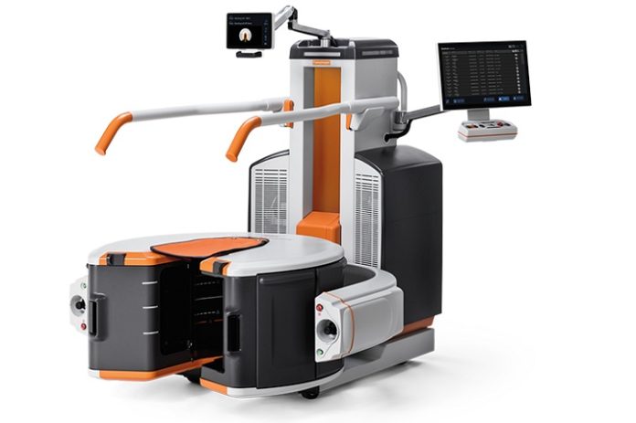
Carestreamโ€™s state-of-the-art OnSight 3D Extremity System will be featured at the upcoming European Congress of Radiology (ECR) meeting in Vienna, Austria (Carestream booth #407, Expo X4). The system enables 3D upper extremity and weight-bearing lower extremity exams, providing affordable and convenient image processing for orthopaedic practices, imaging centers and hospitals at the point of care.

โ€œThe OnSight system delivers high-resolution 3D images with advanced anatomical detail that can help radiologists make more accurate diagnostic decisions and help orthopaedic specialists decide on treatment plans,โ€ said Cyrill Aschenbrenner, Business Director EMEA โ€“ Products & Solutions at Carestream. โ€œIt improves the visibility around anatomical hardware like metal screws, plates or nails in the body.โ€

The CARESTREAM OnSight 3D Extremity System uses cone beam computed tomography (CBCT) technology to capture high-quality images for weight-bearing and other extremity exams that are not possible with traditional CT. Unlike traditional CT systems, the OnSight system has a large-area detector that captures a 3D image of the extremity in a single rotation, which takes only 25 seconds. It employs three X-ray sources to reduce artifacts and improve the overall field of view, capturing the full anatomy of interest in a single scan. Advanced scatter and metal artifact reduction algorithms improve the visibility of patient anatomy and reduce the distracting inยญfluences of metal implants, providing an unobstructed view.

Additionally, the OnSight system can provide high-resolution 3D images that can help reveal subtle or occult fractures and soft-tissue injuries. Surgeons also can study fracture healing over time with accurate images.

โ€œA traditional CT system can be a costly proposition for smaller imaging facilities and orthopaedic practices,โ€ Mr. Aschenbrenner said. โ€œOnSightโ€™s small footprint and simplified design cuts the time and cost of installation, and results in productivity gains for imaging centers and hospitals by freeing up full-body CT systems for other exams.โ€


AI to predict heart attacks and stroke

World first in AI helps scan heart disease patients to predict heart attacks and stroke

Artificial intelligence has been used for the first time to instantly and accurately measure blood flow, in a study led by Barts Health NHS Trust and University College London.

The results were found to be able to predict chances of death, heart attack and stroke, and can be used by doctors to help recommend treatments which could improve a patientโ€™s blood flow.

Heart disease is the leading global cause of death and illness. Reduced blood flow, which is often treatable, is a common symptom of many heart conditions. International guidelines therefore recommend a number of assessments to measure a patientโ€™s blood flow, but many are invasive and carry a risk.

Non-invasive blood flow assessments are available, including cardiovascular magnetic resonance (CMR) imaging, but up until now, the scan images have been incredibly difficult to analyse in a manner precise enough to deliver a prognosis or recommend treatment.

In the largest study of its kind, funded by British Heart Foundation and Barts Charity, and published in the journal Circulation, researchers took routine CMR scans from more than 1,000 patients attending St Bartholomew’s Hospital and the Royal Free Hospital and used a new automated artificial intelligence technique to analyse the images.

By doing this, the teams were able to precisely and instantaneously quantify the blood flow to the heart muscle and deliver the measurements to the medical teams treating the patients.

By comparing the AI-generated blood flow results with the health outcomes of each patient, the team found that the patients with reduced blood flow were more likely to have adverse health outcomes including death, heart attack, stroke and heart failure.

The AI technique was therefore shown for the first time to be able to predict which patients might die or suffer major adverse events, better than a doctor could on their own with traditional approaches.

Professor James Moon from Barts Health NHS Trust and University College London said: โ€œArtificial intelligence is moving out of the computer labs and into the real world of healthcare, carrying out some tasks better than doctors could do alone.

“We have tried to measure blood flow manually before, but it is tedious and time-consuming, taking doctors away from where they are needed most, with their patients.โ€

Dr Kristopher Knott from Barts Health NHS Trust and University College London added: โ€œThe predictive power and reliability of the AI was impressive and easy to implement within a patientโ€™s routine care. “The calculations were happening as the patients were being scanned, and the results were immediately delivered to doctors.

“As poor blood flow is treatable, these better predictions ultimately lead to better patient care, as well as giving us new insights into how the heart works.โ€

Peter Kellman from the National Institutes of Health (NIH) in the United States, who working with Hui Xue at the NIH, developed the automated AI techniques to analyse the images that were used in the study, said: โ€œThis study demonstrates the growing potential of artificial intelligence-assisted imaging technology to improve the detection of heart disease and may move clinicians closer to a precision medicine approach to optimize patient care.

“We hope that this imaging approach can save lives in the future.โ€ Kellman is director of the Medical Signal and Image Processing Program at the National Heart, Lung, and Blood Institute, part of the NIH.

The study was funded by the British Heart Foundation, National Institute for Health Research, European Regional Development Fund and Barts Charity, and involved additional researchers from the Royal Free Hospital, Queen Mary University of London and the University of Leeds.

Mount Sinai Discovers New Drug Combo to Induce High Rates of Human Beta Cell Regeneration

Mount Sinai Discovers New Drug Combo to Induce High Rates of Human Beta Cell Regeneration

Researchers at the Icahn School of Medicine at Mount Sinai have discovered a novel combination of two classes of drugs that, together, cause the highest rate of proliferation ever observed in adult human beta cellsโ€”the cells in the pancreas that produce insulinโ€”without harming most other cells in the body. The result is an important step toward a diabetes treatment that restores the bodyโ€™s ability to produce insulin.

The finding involved one type of drug that is known to cause beta cells to proliferate and another that is already in widespread use in people with diabetes. Together, they caused the cells to proliferate at a rate of 5 to 6 percent per day. The study was published today in Science Translational Medicine online. For a video about this study click here.

โ€œWe are very excited about this new drug combination because for the first time ever, we are able to see rates of human beta cell replication that are sufficient to replenish beta cell mass in humans with diabetes,โ€ said Andrew Stewart, MD, Director of the Mount Sinai Diabetes, Obesity, and Metabolism Institute and lead author of the study.

Diabetes occurs when there are not enough beta cells in the pancreas, or when those beta cells secrete too little insulin, the hormone required to keep blood sugar levels in the normal range. Approximately 30 million people in the United States have diabetes and nearly 50 to 80 million more are living with prediabetes (also called โ€œmetabolic syndromeโ€). Diabetes can lead to major medical complications: heart attack, stroke, kidney failure, blindness, and limb amputation.

In type 1 diabetes, the immune system mistakenly attacks and destroys beta cells. A deficiency of functioning beta cells is also an important contributor to type 2 diabetes, the most common type of diabetes. Thus, developing drugs that can increase the number of healthy beta cells is a major priority in diabetes research.

According to Dr. Stewart, none of the diabetes drugs currently on the market can induce beta cell regeneration in people with diabetes. In parallel with the Mount Sinai work, other researchers are studying pancreatic transplantation, beta cell transplantation, and stem cell replacement of beta cells for people with diabetes, but none of these approaches is in widespread use.

โ€œThis is a very exciting discovery in the field of diabetes and is a key next step in drug development for this disease,โ€ said Dennis S. Charney, MD, Anne and Joel Ehrenkranz Dean, Icahn School of Medicine at Mount Sinai. โ€œThis important work truly holds promise for so many people.โ€

In 2015, Dr. Stewart and his team published a paper in Nature Medicine that showed that harmine, a drug that inhibits the enzyme dual specificity tyrosine-regulated kinase 1A (DYRK1A), induced multiplication of adult human beta cells. In that study, his team also discovered that harmine treatment led to normal control of blood sugar and proliferation in human beta cells in diabetic mice whose beta cells had been replaced with small numbers of transplanted human beta cells. While this was a major advance, the proliferation rate was lower than needed to rapidly expand beta cells in people with diabetes.

This current paper builds upon a study that Dr. Stewart and his team published in Cell Metabolism in December 2018 where they discovered that DYRK1A inhibitors combined with another drug that inhibits transforming growth factor beta superfamily members (TGFฮฒSF), also known as a family of proteins with various biological processes such as growth, development, tissue homeostasis and immune system, could cause beta cells to proliferate at a rate of 5 to 8 percent per day. However, according to Dr. Stewart, TGFฮฒSFโ€™s would likely have side effects on other organs in the body that would prevent clinical use.

The next challenge was developing ways to target regenerative drugs to the beta cells while avoiding other cells and organs in the body where they may elicit adverse effects.

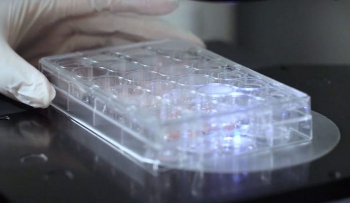
In the study published, titled โ€œGLP-1 receptor agonists synergize with DYRK1A inhibitors to potentiate functional human beta cell regeneration,” Dr. Stewart and his team combined DYRK1A inhibitors like harmine with a class of beta cell-targeting drugs, also known as GLP1R agonists, which are already in widespread use in people with type 2 diabetes. They showedโ€”in beta cells from normal people and people with type 2 diabetes, both in the tissue culture dishes and in human beta cells transplanted into miceโ€”that combining harmine (or any other DYRK1A inhibitor) with any of the many GLP1R agonist drugs currently on the market for diabetes yields high rates of human beta cell replication, and does so in a way that is highly selective for the beta cell.

The project arose from the PhD thesis of an Icahn School of Medicine graduate student, Courtney Ackeifi, now a postdoctoral fellow in Dr. Stewartโ€™s lab and first author of the paper, who explored a broad spectrum of potential drug partners that could enhance the beta cell regenerative efficacy and selectivity of harmine.

Said Dr. Ackeifi of the discovery, โ€œThe beauty here is that the combination of DYRK1A inhibitors with GLP1R agonists achieves the highest rate of human beta cell replication possible, and does so in a highly specific way. This is an important advance in the field of diabetes because we may have found a way to convert a widely used class of diabetes drugs into a potent human beta cell regenerative treatment for all forms of diabetes.โ€

โ€œWe know that a critical pathway to drive a cure for type 1 diabetes includes transplanting insulin-producing beta cells into people or enticing their existing beta cells to start multiplying,โ€ explains Francis Martin, PhD, JDRF Director of Research. โ€œIt is exciting to learn from the work of Dr. Stewart and his team that GLP1R agonists could increase the effect of the recently discovered agents that promote multiplication. Using GLP1R offers a means to boost the effect while also improving the safety of this type of drug.โ€

The next goals of the project are to perform long-term studies in animals transplanted with human beta cells, and to determine if any cells or organs in the body other than beta cells are affected by the new drug combination.

This work was supported by funding from: NIH grants T32 GM062754 (to CA), DK105015 (to AFS), DK105015-01A1S1 (to CA), P-30 DK 020541 (to AFS and AG-O) and UC4 DK104211 (to AFS), by JDRF Grants 2-SRA- 2015-62 (to AFS) and 2-SRA-2017 514-S-B (to AFS), and by the Icahn School of Medicine at Mount Sinai.

Also making key research contributions were Esra Karakose, PhD; Hongtao Liu, BS; Jessica Wilson, BS; Ethan Swartz, BS; Cecilia Berroute, BS; Yansui Li, MD; Kunal Kumar, PhD; Roberto Sanchez, PhD; Robert DeVita, PhD; Dirk Homann, MD, PhD; Donald K. Scott, PhD; and Adolfo Garcia-Ocaรฑa, PhD from the Diabetes, Obesity, and Metabolism Institute, the Department of Pharmacological Sciences, and The Drug Discovery Institute at the Icahn School of Medicine at Mount Sinai.

Additional investigators included researchers from The Naomi Berrie Diabetes Center and the Columbia Stem Cell Center at Columbia University (Bryan J. Gonzรกlez, BS; Dieter Egli, PhD); The Department of Pharmacology and The Alberta Diabetes Institute, The University of Alberta, Edmonton, Alberta, Canada (Jocelyn E. Manning Fox, PhD; Patrick E. MacDonald, PhD); and the Center for Integrative Genomics at the University of Lausanne in Switzerland (Bernard Thorens, PhD).

About JDRF

JDRF is the leading global organization funding type 1 diabetes (T1D) research. Our mission is to accelerate life-changing breakthroughs to cure, prevent and treat T1D and its complications. To accomplish this, JDRF has invested more than $2.2 billion in research funding since our inception. We are an organization built on a grassroots model of people connecting in their local communities, collaborating regionally for efficiency and broader fundraising impact, and uniting on a national stage to pool resources, passion, and energy. We collaborate with academic institutions, policymakers, and corporate and industry partners to develop and deliver a pipeline of innovative therapies to people living with T1D. Our staff and volunteers throughout the United States and our six international affiliates are dedicated to advocacy, community engagement and our vision of a world without T1D.

About the Mount Sinai Health System

The Mount Sinai Health System is New York City’s largest academic medical system, encompassing eight hospitals, a leading medical school, and a vast network of ambulatory practices throughout the greater New York region. Mount Sinai is a national and international source of unrivaled education, translational research and discovery, and collaborative clinical leadership ensuring that we deliver the highest quality careโ€”from prevention to treatment of the most serious and complex human diseases. The Health System includes more than 7,200 physicians and features a robust and continually expanding network of multispecialty services, including more than 400 ambulatory practice locations throughout the five boroughs of New York City, Westchester, and Long Island. The Mount Sinai Hospital is ranked No. 14 on U.S. News & World Report’s “Honor Roll” of the Top 20 Best Hospitals in the country and the Icahn School of Medicine as one of the Top 20 Best Medical Schools in country. Mount Sinai Health System hospitals are consistently ranked regionally by specialty and our physicians in the top 1% of all physicians nationally by U.S. News & World Report.


 

Much shorter radiation treatment found to be safe, effective for people with soft tissue sarcoma

Much shorter radiation treatment found to be safe, effective for people with soft tissue sarcoma

A new study led by researchers at the UCLA Jonsson Comprehensive Cancer Center found that treating soft tissue sarcoma with radiation over a significantly shorter period of time is safe, and likely just as effective, as a much longer conventional course of treatment.

Sarcoma, a rare type of cancer of the soft tissues (for example, muscle, nerves, fat, or fibrous tissue) or bone, affects about 13,000 people of all ages in the United States each year. Many people with a soft tissue sarcoma diagnosis receive a five-week course of radiation therapy (daily Monday through Friday). Four to five weeks after that, the tumor is removed in surgery. In this study at UCLA, a national leader in the care of patients with sarcoma, researchers used a condensed five-day radiation regimen that considerably cuts down the length of treatment and the time to surgery.

“Shortening the radiation therapy from five weeks to five days has been a very meaningful change for patients,” said lead author Dr. Anusha Kalbasi, assistant professor of radiation oncology in the division of molecular and cellular oncology. “Five weeks of daily treatments is a burdensome commitment for patients. The daily back-and-forth can be expensive and time consuming, and it can really interfere with work, school or parenting. So finding a way to safely shorten the radiation treatment is a significant advancement in improving the quality of care for patients with hard-to-treat cancers like sarcoma.”

Researchers enrolled 52 adults at UCLA diagnosed with a soft tissue sarcoma of the limbs or trunk โ€” where these tumors most commonly occur โ€” that was localized and had not spread to other parts of the body. The participants underwent a condensed form of radiation therapy for five days, followed by surgery. The team then analyzed and followed the cohort for an average of 2 1/2 years.

They found less than 6% of the patients with at least two years of follow-up on the clinical trial had a recurrence of their tumor, which is on par with studies using the conventional five-week regimen, Kalbasi said.

The main reason we treat sarcoma patients with radiation before surgery is to prevent the tumor from recurring where it was removed,” said Kalbasi, who is also a member of the Jonsson Cancer Center. “As an added benefit, in some cases it can cause the tumor to shrink. So far it appears that the five-day treatment is working just as well as the five-week treatment.”

Along with studying tumor recurrence rates, researchers also closely examined the severity of wound complications. Sarcoma surgery often requires a large incision to remove the tumor and can by itself result in wound complications. Adding radiation before surgery can slow the healing process even more. The team wanted to make sure the condensed five-day treatment didn’t make wound complications worse.

“In the long term, having radiation before surgery lowers the chance of side effects like swelling, joint stiffness and fibrosis, that are big contributors to quality of life,” Kalbasi said. “But this comes at a trade-off with a higher rate of wound complications immediately after surgery. The good news is that our five-day treatment did not increase the chance for wound issues after surgery compared to the conventional five-week treatment.”

Not only did the researchers find that wound complications were virtually the same for the two therapies, but they also identified a new set of biomarkers that could indicate how likely someone will be to encounter significant wound complications.

By using a DNA swab from each participant to test for mutations, the team found that they could identify microRNA-disrupting genetic biomarkers that are associated with the risk of wound complications.

“We know from experience that different patients respond to the same amount of radiation in different ways, and we have always believed that it is due to the patient’s personal response to radiation,” said study co-author Dr. Joanne Weidhaas, a professor of radiation oncology and director of translational research at the David Geffen School of Medicine at UCLA, who led the biomarker studies. “This type of biomarker lets us know up front who may or may not have a high risk of wound complications, and can potentially help us determine the best type of treatment to offer for patients. Having this information upfront is a big advantage, which can really advance personalized medicine.”

In the two years since the study began, the team also noticed a rapid rise in the number of patients coming to UCLA, a high-volume sarcoma center, for radiation treatment before surgery. Nearly half of the study participants traveled more than 100 miles to UCLA for radiation treatment. Past studies have shown treating sarcomas at a high-volume center with the expertise of a multidisciplinary sarcoma team is associated with improved outcomes.

The five-day radiation therapy regimen may not only preferred by people undergoing the treatment, but it will also result in more people having access to care at high-volume centers that specialize in treating such a rare cancer, Kalbasi said.

To further validate the study’s findings, the team plans to continue to follow the cohort for at least five years to see if, in terms of cancer control and longer-term side effects, the five-day radiation treatment continues to be just as safe and effective as the traditional five-week treatment. The team has also expanded the clinical trial to include patients who are also receiving chemotherapy before surgery.

The study was published in Clinical Cancer Research, a journal of the American Association for Cancer Research.

The study’s senior author is Dr. Fritz Eilber, professor of surgical oncology and molecular and medical pharmacology at the Geffen School and director of the UCLA Jonsson Comprehensive Cancer Center Sarcoma Program.

LABORIE Medical Technologies Announces Completion of Clinical Innovations Acquisition

LABORIE Medical Technologies Announces Completion of Clinical Innovations Acquisition

LABORIE Medical Technologies, a leading diagnostic and therapeutic medical technology company, announced it has completed its acquisition of Clinical Innovations for an enterprise value of $525 million.

Based in Salt Lake City, Clinical Innovations is a leading global provider of clinician-preferred medical devices for the labor & delivery (L&D) and neonatal intensive care unit (NICU) specialties.

โ€œWe are pleased to welcome the incredibly talented Clinical Innovations team to the LABORIE family,โ€ commented Michael Frazzette, President & CEO of LABORIE Medical Technologies. โ€œWe look forward to investing in, and building upon Clinical Innovationsโ€™ unique product portfolio and specialty channels to continue to benefit mothers, babies and healthcare professionals around the world.โ€

In order to drive the focus, resources, investments and customer-intense culture needed to continue to be successful in the L&D and NICU specialties, the Clinical Innovations business will form a stand-alone Global Business Unit (GBU) called Maternal and Child Health.

โ€œThis focused GBU approach will enable us to build on Clinical Innovationsโ€™ established strong and trusted relationships with physicians and nurses; and serve the distinct customer call point of obstetricians, gynecologists, neonatologists, and NICU and L&D nurses,โ€ Frazzette added.

โ€œWe look forward to leveraging Patricia Industriesโ€™ healthcare expertise and global presence to support the combined LABORIE and Clinical Innovations organization as we embark on our next phase of growth together,” said Yuriy Prilutskiy, Managing Director at Patricia Industries, a part of Investor AB, and owner of LABORIE Medical Technologies. โ€œConsistent with our purpose of creating value for people and society by building strong and sustainable businesses, we will continue to invest in and develop the company to improve clinical outcomes for patients around the world for many years to come.โ€

Piper Jaffray acted as financial advisor and Cleary Gottlieb Steen & Hamilton LLP as legal advisor to LABORIE.

GenesisCare officially opens new cancer care centre with capacity to treat 500 patients a year

GenesisCare officially opens new cancer care centre with capacity to treat 500 patients a year

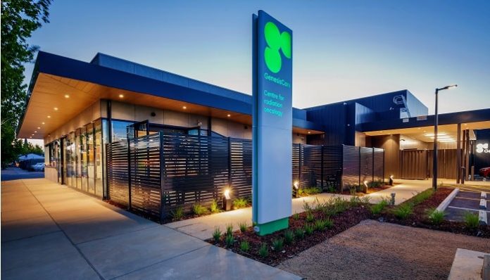
For the first time, cancer patients in Goulburn Valley no longer have to travel outside the local community for treatment, thanks to a new $11 million cancer care centre unveiled at an official opening ceremony.

The cancer treatment centre, which began delivering care to patients before Christmas, is open to both public and privately insured patients, and has capacity to treat more than 500 patients every year.

In December 2019, the Department of Health and Human Services, Goulburn Valley Health and GenesisCare, announced a shared care agreement to enable public patient access to treatment at the GenesisCare Shepparton centre. All patients at the centre can receive world-class cancer treatments and access to clinical trials, closer to home.

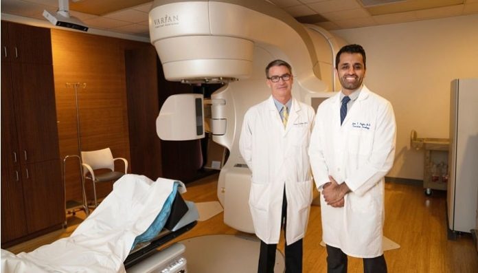
The facility provides comprehensive radiation therapy services using an advanced Varian TrueBeam linear accelerator machine to deliver highly targeted doses of radiation to tumours using Volumetric Modulated Arc Therapy (VMAT). International guidelines recommend radiation therapy in the treatment of half of all cancer cases.

Previously, patients had to commute hundreds of kilometres to Albury, Bendigo or Melbourne for daily radiation therapy treatment or be away from home for multiple weeks. A local service opens up access for many patients, including those who would benefit from rapid access to radiation therapy for pain management as part of their palliative care. Additional cutting-edge treatment techniques will be introduced at the centre in 2020.

The Hon Damian Drum, Federal Member for Murray, said: โ€œThe official opening of the new centre is a huge step forward for cancer care in the Goulburn Valley region and for regional Victoria. Previously, local patients were travelling up to 200kms from Shepparton to Melbourne, Albury or Bendigo for treatment, so this is fantastic news for the local community.

โ€œThe Federal Government is very proud to have assisted with the purchase of the advanced linear accelerator machine, which is now being used to deliver radiation therapy for local patients.โ€

Shane Ryan, GenesisCare Victoria Oncology General Manager, said: โ€œAt GenesisCare, we are committed to ensuring all cancer patients, regardless of where they live, receive access to the highest standards of care close to home.

โ€œWe are pleased to be up and running, and to be partnering alongside the local community, the Department of Health and Goulburn Valley Health to deliver world-class treatment.โ€

A team of more 12 experienced and respected oncologists, nurses and support staff have been recruited to deliver the highest quality of cancer care for the local community. They are supported by an international network of more than 3000 healthcare professions, including leading specialists across every tumour type.

โ€œA special mention must also go to our fantastic local trades teams who worked tirelessly to ensure we could start offering treatment to patients as soon as possible.โ€

Dr Grace Ong, GenesisCare Shepparton Radiation Oncologist, said: โ€œThe entire team here at GenesisCare in Shepparton is incredibly passionate about improving the health outcomes of people living in the Goulburn Valley.

โ€œWe have treated more than 50 patients since Christmas and the feedback has been overwhelmingly positive. The whole community has embraced our team with open arms, and they are just so pleased they no longer have to travel for treatment.โ€

The facility is located at 124 Corio Street in the centre of the town, opposite GVH Community Health. No private health cover is required. For private patients, the cost of radiation therapy treatment is largely covered through Medicare via the Medicare Benefits Scheme and the Extended Medicare Safety Net programs.


CloudCath raises funds towards peritoneal dialysis monitoring system

CloudCath raises funds towards peritoneal dialysis monitoring system

Digital health medical device company CloudCath has completed a $12m Series A funding round to commercialise a system that enables remote and live monitoring for patients undergoing at-home peritoneal dialysis.

It also plans to use the funds to support the development of additional applications utilising the companyโ€™s proprietary technology. Capital Integral led the funding round, with participation from FundRx, The Capital Partnership, Coconut Tree Investments, Stanford Universityโ€™s StartX Fund and additional strategic and individual investors.

Seamlessly integrating into the patientโ€™s drain line, CloudCathโ€™s device helps to monitor the spent dialysate fluid on every dialysis cycle by measuring and quantifying the fluid properties remotely from the patientsโ€™ home.

CloudCathโ€™s monitoring platform helps healthcare providers to log in and access real-time data from patients throughout treatment, to assist in decision management. CloudCath co-founder and CEO Aly ElBadr said: โ€œEnd-stage renal disease, one of the costliest chronic diseases in the US, is devastating to patients.

โ€œThe CloudCath technology enables patients and caregivers to easily monitor fluid parameters for early signs of complications, including infection, in real-time, allowing dialysis providers to intervene much earlier than the existing standard of care.

Translate ยป