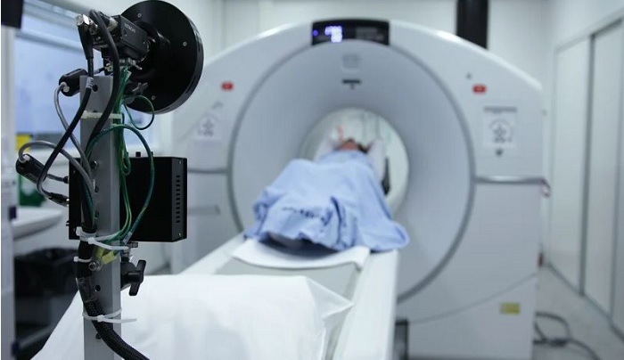
Dynamic contrast-enhanced MRI can help physicians detect facial nerve abnormalities commonly seen in people with Bell’s palsy, researchers reported. In patients with BP, facial nerves become inflamed or swollen causing temporary weakness or partial paralysis of facial muscles, Chinese researchers explained in Clinical Radiology.
And while DCE has been proven to enhance traditional MR imaging, due to inherent limitations with MRI, specialists have tried to use a number of different sequences to accurately spot facial features consistent with BP.
“On the one hand, some segments of the facial nerve are too small to be assessed on conventional MRI,” Y. Wang, a radiologist with Shanghai Ninth People’s Hospital, and colleagues commented. “On the other hand, a normal facial nerve has a certain probability of presenting enhancement on conventional MRI.”
The authors sought to test if DCE-MRI could do the job, retrospectively analyzing 13 patients with surgically confirmed Bell’s palsy. Exams were completed using a T1-weighted volumetric interpolated breath-hold examination (VIBE) sequence between January 2015 and July 2019.
They determined that DCE-MRI was more accurate at imaging the involved segments of BP-affected facial nerves than conventional MR imaging (92.3% vs. 38.5%).
Additionally, Wang and colleagues found the VIBE sequence produced images with better contrast, higher signal-to-noise ratios, and a shorter exam time.
While DCE-MRI proved valuable, the approach should still be considered a complementary tool to morphological imaging, the researchers noted.
“In conclusion, conventional MRI combined with DCE-MRI is a useful way to diagnose the involved segments of the affected facial nerves accurately with a shorter acquisition time compared to conventional MRI.
This approach has advantages both for the patient, in terms of safety, and for the physician, in terms of the accuracy of the diagnosis,” the group concluded.


